


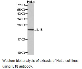
Figure 1. Western blot analysis of IL18 using anti-IL18 antibody (PB9006). Electrophoresis was performed on a 5-20% SDS-PAGE gel at 70V (Stacking gel) / 90V (Resolving gel) for 2-3 hours. The sample well of each lane was loaded with 30 ug of sample under reducing conditions. Lane 1: human Hela whole cell lysates, Lane 2: human HepG2 whole cell lysates. After electrophoresis, proteins were transferred to a nitrocellulose membrane at 150 mA for 50-90 minutes. Blocked the membrane with 5% non-fat milk/TBS for 1.5 hour at RT. The membrane was incubated with rabbit anti-IL18 antigen affinity purified polyclonal antibody (Catalog # PB9006) at 0.5 microg/mL overnight at 4°C, then washed with TBS-0.1%Tween 3 times with 5 minutes each and probed with a goat anti-rabbit IgG-HRP secondary antibody at a dilution of 1:5000 for 1.5 hour at RT. The signal is developed using an Enhanced Chemiluminescent detection (ECL) kit (Catalog # EK1002) with Tanon 5200 system. A specific band was detected for IL18 at approximately 22 kDa. The expected band size for IL18 is at 22 kDa.
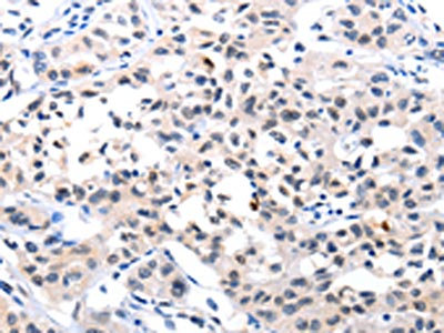
. IL18 was detected in a paraffin-embedded section of human tonsil tissue. Heat mediated antigen retrieval was performed in EDTA buffer (pH 8.0, epitope retrieval solution). The tissue section was blocked with 10% goat serum. The tissue section was then incubated with 2 microg/ml rabbit anti-IL18 Antibody (PB9006) overnight at 4°C. Peroxidase Conjugated Goat Anti-rabbit IgG was used as secondary antibody and incubated for 30 minutes at 37°C. The tissue section was developed using HRP Conjugated Rabbit IgG Super Vision Assay Kit (Catalog # SV0002) with DAB as the chromogen.")
 and anti-Beta Tubulin antibody (M01857-3). IL18 was detected in immunocytochemical section of A549 cell. Enzyme antigen retrieval was performed using IHC enzyme antigen retrieval reagent (AR0022) for 15 mins. The cells were blocked with 10% goat serum. And then incubated with 5 microg/mL rabbit anti-IL18 Antibody (PB9006) and mouse anti-Beta Tubulin antibody (M01857-3) overnight at 4°C. Cy3 Conjugated Goat Anti-Rabbit IgG (BA1032) and DyLight®488 Conjugated Goat Anti-Mouse IgG (BA1126) were used as secondary antibody at 1:500 dilution and incubated for 30 minutes at 37°C. Visualize using a fluorescence microscope and filter sets appropriate for the label used.")
Figure 1. Western blot analysis of IL18 using anti-IL18 antibody (PB9006). Electrophoresis was performed on a 5-20% SDS-PAGE gel at 70V (Stacking gel) / 90V (Resolving gel) for 2-3 hours. The sample well of each lane was loaded with 30 ug of sample under reducing conditions. Lane 1: human Hela whole cell lysates, Lane 2: human HepG2 whole cell lysates. After electrophoresis, proteins were transferred to a nitrocellulose membrane at 150 mA for 50-90 minutes. Blocked the membrane with 5% non-fat milk/TBS for 1.5 hour at RT. The membrane was incubated with rabbit anti-IL18 antigen affinity purified polyclonal antibody (Catalog # PB9006) at 0.5 microg/mL overnight at 4°C, then washed with TBS-0.1%Tween 3 times with 5 minutes each and probed with a goat anti-rabbit IgG-HRP secondary antibody at a dilution of 1:5000 for 1.5 hour at RT. The signal is developed using an Enhanced Chemiluminescent detection (ECL) kit (Catalog # EK1002) with Tanon 5200 system. A specific band was detected for IL18 at approximately 22 kDa. The expected band size for IL18 is at 22 kDa.
Anti-IL18 Antibody Picoband(r)
PB9006-CARRIER-FREE
ApplicationsImmunoFluorescence, Western Blot, ImmunoCytoChemistry, ImmunoHistoChemistry
Product group Antibodies
ReactivityHuman
TargetIL18
Overview
- SupplierBoster Bio
- Product NameAnti-IL18 Antibody Picoband(r)
- Delivery Days Customer9
- Application Supplier NoteWB: The detection limit for IL-18 is approximately 0.25ng/lane under reducing conditions. Tested Species: In-house tested species with positive results. By Heat: Boiling the paraffin sections in 10mM citrate buffer, pH6.0, for 20mins is required for the staining of formalin/paraffin sections. Other applications have not been tested. Optimal dilutions should be determined by end users.
- ApplicationsImmunoFluorescence, Western Blot, ImmunoCytoChemistry, ImmunoHistoChemistry
- CertificationResearch Use Only
- ClonalityPolyclonal
- Concentration500 ug/ml
- Gene ID3606
- Target nameIL18
- Target descriptioninterleukin 18
- Target synonymsIGIF, IL-18, IL-1g, IL1F4, interleukin-18, IFN-gamma-inducing factor, IL-1 gamma, iboctadekin, interleukin 18 (interferon-gamma-inducing factor), interleukin-1 gamma
- HostRabbit
- IsotypeIgG
- Protein IDQ14116
- Protein NameInterleukin-18
- Scientific DescriptionBoster Bio Anti-IL18 Antibody Picoband® catalog # PB9006. Tested in IF, IHC, ICC, WB applications. This antibody reacts with Human. The brand Picoband indicates this is a premium antibody that guarantees superior quality, high affinity, and strong signals with minimal background in Western blot applications. Only our best-performing antibodies are designated as Picoband, ensuring unmatched performance.
- ReactivityHuman
- Storage Instruction-20°C,2°C to 8°C
- UNSPSC12352203
Related products
Product group Antibodies
IL18 AntibodyCSB-PA081015
ApplicationsWestern Blot, ELISA, ImmunoHistoChemistry
ReactivityHuman
TargetIL18
- SizePrice
Product group Antibodies

Anti-IL-18 [125-2H]Ab00140-1.1
ApplicationsImmunoFluorescence, ImmunoPrecipitation, ELISA, Neutralisation/Blocking
ReactivityHuman
TargetIL18
- SizePrice
Product group Antibodies
Anti-IL18 AntibodyA30073
ApplicationsWestern Blot, ImmunoHistoChemistry
ReactivityHuman
- SizePrice
Product group Antibodies

Goat anti-IL18EB09393
ApplicationsFlow Cytometry, ImmunoFluorescence, Western Blot, ELISA
ReactivityHuman
TargetIL18
- SizePrice

Product group Antibodies
Anti-IL18 AntibodyHPA003980
ApplicationsWestern Blot, ImmunoCytoChemistry, ImmunoHistoChemistry
ReactivityHuman
TargetIL18
- SizePrice
Product group Antibodies
IL18 Polyclonal Antibodybs-4988R
ApplicationsImmunoFluorescence, Western Blot, ImmunoHistoChemistry, ImmunoHistoChemistry Paraffin
ReactivityHuman
TargetIL18
- SizePrice
Product group Antibodies

IL18 AntibodyLS-C404072
ApplicationsWestern Blot, ELISA, ImmunoHistoChemistry
ReactivityHuman
TargetIL18
- SizePrice
Product group Antibodies
IL18 Polyclonal AntibodyCAC13342
ApplicationsELISA, ImmunoHistoChemistry
TargetIL18
- SizePrice
![IL18 antibody [HL1761] detects IL18 protein at cytoplasm by immunohistochemical analysis. Sample: Paraffin-embedded human tonsil. IL18 stained by IL18 antibody [HL1761] (GTX637411) diluted at 1:100. Antigen Retrieval: Citrate buffer, pH 6.0, 15 min](https://www.genetex.com/upload/website/prouct_img/normal/GTX637411/GTX637411_T-44788_20221007_IHC-P_22110201_896.webp)
Product group Antibodies
IL18 antibody [HL1761]GTX637411
ApplicationsWestern Blot, ELISA, ImmunoHistoChemistry, ImmunoHistoChemistry Paraffin
ReactivityHuman
TargetIL18
- SizePrice