


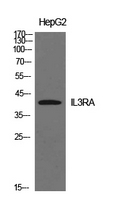
Figure 1. Western blot analysis of IL3RA using anti-IL3RA antibody (A04307). Electrophoresis was performed on a 5-20% SDS-PAGE gel at 70V (Stacking gel) / 90V (Resolving gel) for 2-3 hours. The sample well of each lane was loaded with 50ug of sample under reducing conditions. Lane 1: human Raji whole cell lysates, Lane 2: human K562 whole cell lysates. After Electrophoresis, proteins were transferred to a Nitrocellulose membrane at 150mA for 50-90 minutes. Blocked the membrane with 5% Non-fat Milk/ TBS for 1.5 hour at RT. The membrane was incubated with rabbit anti-IL3RA antigen affinity purified polyclonal antibody (Catalog # A04307) at 0.5 microg/mL overnight at 4°C, then washed with TBS-0.1%Tween 3 times with 5 minutes each and probed with a goat anti-rabbit IgG-HRP secondary antibody at a dilution of 1:5000 for 1.5 hour at RT. The signal is developed using an Enhanced Chemiluminescent detection (ECL) kit (Catalog # EK1002) with Tanon 5200 system. A specific band was detected for IL3RA at approximately 60KD. The expected band size for IL3RA is at 60KD.
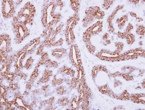
. IL3RA was detected in paraffin-embedded section of human rectal cancer tissue. Heat mediated antigen retrieval was performed in EDTA buffer (pH8.0, epitope retrieval solution). The tissue section was blocked with 10% goat serum. The tissue section was then incubated with 1microg/ml rabbit anti-IL3RA Antibody (A04307) overnight at 4°C. Biotinylated goat anti-rabbit IgG was used as secondary antibody and incubated for 30 minutes at 37°C. The tissue section was developed using Strepavidin-Biotin-Complex (SABC) (Catalog # SA1022) with DAB as the chromogen.")
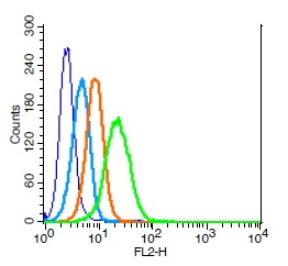
. Overlay histogram showing Raji cells stained with A04307 (Blue line). The cells were fixed with 4% paraformaldehyde and blocked with 10% normal goat serum. And then incubated with rabbit anti-IL3RA Antibody (A04307, 1microg/1x106 cells) for 30 min at 20°C. DyLight®488 conjugated goat anti-rabbit IgG (BA1127, 5-10microg/1x106 cells) was used as secondary antibody for 30 minutes at 20°C. Isotype control antibody (Green line) was rabbit IgG (1microg/1x106) used under the same conditions. Unlabelled sample without incubation with primary antibody and secondary antibody (Red line) was used as a blank control.")
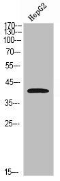
Figure 1. Western blot analysis of IL3RA using anti-IL3RA antibody (A04307). Electrophoresis was performed on a 5-20% SDS-PAGE gel at 70V (Stacking gel) / 90V (Resolving gel) for 2-3 hours. The sample well of each lane was loaded with 50ug of sample under reducing conditions. Lane 1: human Raji whole cell lysates, Lane 2: human K562 whole cell lysates. After Electrophoresis, proteins were transferred to a Nitrocellulose membrane at 150mA for 50-90 minutes. Blocked the membrane with 5% Non-fat Milk/ TBS for 1.5 hour at RT. The membrane was incubated with rabbit anti-IL3RA antigen affinity purified polyclonal antibody (Catalog # A04307) at 0.5 microg/mL overnight at 4°C, then washed with TBS-0.1%Tween 3 times with 5 minutes each and probed with a goat anti-rabbit IgG-HRP secondary antibody at a dilution of 1:5000 for 1.5 hour at RT. The signal is developed using an Enhanced Chemiluminescent detection (ECL) kit (Catalog # EK1002) with Tanon 5200 system. A specific band was detected for IL3RA at approximately 60KD. The expected band size for IL3RA is at 60KD.
Anti-IL3RA/CD123 Antibody Picoband(r)
A04307-CARRIER-FREE
ApplicationsFlow Cytometry, Western Blot, ELISA, ImmunoHistoChemistry
Product group Antibodies
ReactivityHuman
TargetIL3RA
Overview
- SupplierBoster Bio
- Product NameAnti-IL3RA/CD123 Antibody Picoband(r)
- Delivery Days Customer9
- Application Supplier NoteTested Species: In-house tested species with positive results. Other applications have not been tested. Optimal dilutions should be determined by end users.
- ApplicationsFlow Cytometry, Western Blot, ELISA, ImmunoHistoChemistry
- CertificationResearch Use Only
- ClonalityPolyclonal
- Concentration500 ug/ml
- Gene ID3563
- Target nameIL3RA
- Target descriptioninterleukin 3 receptor subunit alpha
- Target synonymsCD123, IL-3R-alpha, IL3R, IL3RAY, IL3RX, IL3RY, hIL-3Ra, interleukin-3 receptor subunit alpha, CD123 antigen, IL-3 receptor subunit alpha, IL-3R subunit alpha, interleukin 3 receptor, alpha (low affinity)
- HostRabbit
- IsotypeIgG
- Protein IDP26951
- Protein NameInterleukin-3 receptor subunit alpha
- Scientific DescriptionBoster Bio Anti-IL3RA/CD123 Antibody Picoband® catalog # A04307. Tested in ELISA, Flow Cytometry, IHC, WB applications. This antibody reacts with Human. The brand Picoband indicates this is a premium antibody that guarantees superior quality, high affinity, and strong signals with minimal background in Western blot applications. Only our best-performing antibodies are designated as Picoband, ensuring unmatched performance.
- ReactivityHuman
- Storage Instruction-20°C,2°C to 8°C
- UNSPSC12352203
Related products
Product group Antibodies

Anti-CD123 [7G3]Ab02242-10.0
ApplicationsFlow Cytometry, ImmunoPrecipitation, Western Blot, Neutralisation/Blocking
ReactivityHuman
TargetIL3RA
- SizePrice
Product group Antibodies
Anti-IL3RA AntibodyA101017
ApplicationsWestern Blot, ELISA
ReactivityHuman
- SizePrice
Product group Antibodies
Anti-IL3RA Antibody144-03926
ApplicationsWestern Blot
ReactivityHuman, Mouse, Rat
TargetIL3RA
- SizePrice
Product group Antibodies
IL3RA Polyclonal AntibodyBS-2600R
ApplicationsFlow Cytometry, ImmunoFluorescence, Western Blot, ELISA, ImmunoCytoChemistry, ImmunoHistoChemistry, ImmunoHistoChemistry Frozen, ImmunoHistoChemistry Paraffin
ReactivityHuman, Mouse
TargetIL3RA
- SizePrice
Product group Antibodies
IL3RA AntibodyCSB-PA006148
ApplicationsWestern Blot, ELISA
ReactivityHuman
TargetIL3RA
- SizePrice
Product group Antibodies

Goat anti-IL3RA / CD123EB12522
ApplicationsWestern Blot, ELISA, ImmunoHistoChemistry
ReactivityHuman
TargetIL3RA
- SizePrice
Product group Antibodies
ApplicationsFlow Cytometry
TargetIL3RA
- SizePrice
Product group Antibodies

IL3RA / CD123 Antibody (clone 6H6)LS-C106863
ApplicationsFlow Cytometry, ImmunoHistoChemistry, ImmunoHistoChemistry Frozen, ImmunoHistoChemistry Paraffin
ReactivityHuman
TargetIL3RA
- SizePrice
Product group Antibodies
ApplicationsWestern Blot, ImmunoHistoChemistry, ImmunoHistoChemistry Paraffin
ReactivityHuman
TargetIL3RA
- SizePrice