

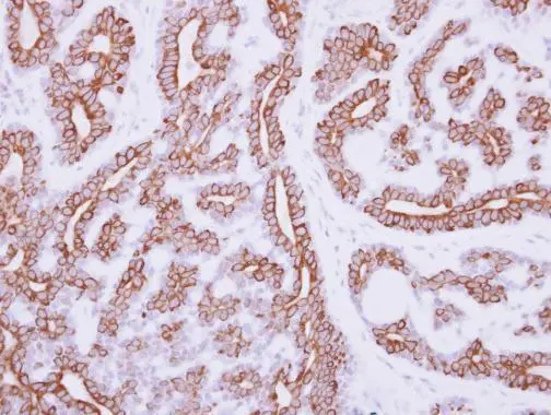
Immunohistochemical analysis of paraffin-embedded human breast cancer, using IL3 Receptor alpha(GTX101563) antibody at 1:250 dilution.
Antigen Retrieval: Trilogy? (EDTA based, pH 8.0) buffer, 15min
, non-transfected (–) and transfected (+) 293T whole cell extracts (5 μg) were separated by 10% SDS-PAGE, and the membrane was blotted with Estrogen Receptor beta antibody (GTX101563) diluted at 1:1000.")
![IL3 Receptor alpha antibody [N2C2], Internal detects IL3 Receptor alpha protein at cell membrane and cytoplasm in human esophageal cancer by immunohistochemical analysis. Sample: Paraffin-embedded human esophageal cancer. IL3 Receptor alpha antibody [N2C2], Internal (GTX101563) diluted at 1:500.
Antigen Retrieval: Citrate buffer, pH 6.0, 15 min](https://www.genetex.com/upload/website/prouct_img/normal/GTX101563/GTX101563_42214_20151012_IHC-P_w_23060100_655.webp "IL3 Receptor alpha antibody [N2C2], Internal detects IL3 Receptor alpha protein at cell membrane and cytoplasm in human esophageal cancer by immunohistochemical analysis. Sample: Paraffin-embedded human esophageal cancer. IL3 Receptor alpha antibody [N2C2], Internal (GTX101563) diluted at 1:500.
Antigen Retrieval: Citrate buffer, pH 6.0, 15 min")
Immunohistochemical analysis of paraffin-embedded human breast cancer, using IL3 Receptor alpha(GTX101563) antibody at 1:250 dilution.
Antigen Retrieval: Trilogy? (EDTA based, pH 8.0) buffer, 15min
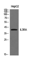
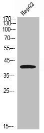
IL3 Receptor alpha antibody [N2C2], Internal
GTX101563
ApplicationsWestern Blot, ImmunoHistoChemistry, ImmunoHistoChemistry Paraffin
Product group Antibodies
ReactivityHuman
TargetIL3RA
Overview
- SupplierGeneTex
- Product NameIL3 Receptor alpha antibody [N2C2], Internal
- Delivery Days Customer9
- Application Supplier NoteWB: 1:500-1:3000. IHC-P: 1:100-1:1000. *Optimal dilutions/concentrations should be determined by the researcher.Not tested in other applications.
- ApplicationsWestern Blot, ImmunoHistoChemistry, ImmunoHistoChemistry Paraffin
- CertificationResearch Use Only
- ClonalityPolyclonal
- Concentration1.2 mg/ml
- ConjugateUnconjugated
- Gene ID3563
- Target nameIL3RA
- Target descriptioninterleukin 3 receptor subunit alpha
- Target synonymsCD123, IL-3R-alpha, IL3R, IL3RAY, IL3RX, IL3RY, hIL-3Ra, interleukin-3 receptor subunit alpha, CD123 antigen, IL-3 receptor subunit alpha, IL-3R subunit alpha, interleukin 3 receptor, alpha (low affinity)
- HostRabbit
- IsotypeIgG
- Protein IDP26951
- Protein NameInterleukin-3 receptor subunit alpha
- Scientific DescriptionThe protein encoded by this gene is an interleukin 3 specific subunit of a heterodimeric cytokine receptor. The receptor is comprised of a ligand specific alpha subunit and a signal transducing beta subunit shared by the receptors for interleukin 3 (IL3), colony stimulating factor 2 (CSF2/GM-CSF), and interleukin 5 (IL5). The binding of this protein to IL3 depends on the beta subunit. The beta subunit is activated by the ligand binding, and is required for the biological activities of IL3. This gene and the gene encoding the colony stimulating factor 2 receptor alpha chain (CSF2RA) form a cytokine receptor gene cluster in a X-Y pseudoautosomal region on chromosomes X or Y. [provided by RefSeq]
- ReactivityHuman
- Storage Instruction-20°C or -80°C,2°C to 8°C
- UNSPSC41116161
Datasheet
Related products
Product group Antibodies

Anti-CD123 [7G3]Ab02242-10.0
ApplicationsFlow Cytometry, ImmunoPrecipitation, Western Blot, Neutralisation/Blocking
ReactivityHuman
TargetIL3RA
- SizePrice
Product group Antibodies
Anti-IL3RA AntibodyA101017
ApplicationsWestern Blot, ELISA
ReactivityHuman
- SizePrice

Product group Antibodies
Anti-IL3RA/CD123 Antibody Picoband(r)A04307-CARRIER-FREE
ApplicationsFlow Cytometry, Western Blot, ELISA, ImmunoHistoChemistry
ReactivityHuman
TargetIL3RA
- SizePrice
Product group Antibodies
Anti-IL3RA Antibody144-03926
ApplicationsWestern Blot
ReactivityHuman, Mouse, Rat
TargetIL3RA
- SizePrice
Product group Antibodies
IL3RA Polyclonal AntibodyBS-2600R
ApplicationsFlow Cytometry, ImmunoFluorescence, Western Blot, ELISA, ImmunoCytoChemistry, ImmunoHistoChemistry, ImmunoHistoChemistry Frozen, ImmunoHistoChemistry Paraffin
ReactivityHuman, Mouse
TargetIL3RA
- SizePrice
Product group Antibodies
IL3RA AntibodyCSB-PA006148
ApplicationsWestern Blot, ELISA
ReactivityHuman
TargetIL3RA
- SizePrice
Product group Antibodies

Goat anti-IL3RA / CD123EB12522
ApplicationsWestern Blot, ELISA, ImmunoHistoChemistry
ReactivityHuman
TargetIL3RA
- SizePrice
Product group Antibodies
ApplicationsFlow Cytometry
TargetIL3RA
- SizePrice
Product group Antibodies

IL3RA / CD123 Antibody (clone 6H6)LS-C106863
ApplicationsFlow Cytometry, ImmunoHistoChemistry, ImmunoHistoChemistry Frozen, ImmunoHistoChemistry Paraffin
ReactivityHuman
TargetIL3RA
- SizePrice