

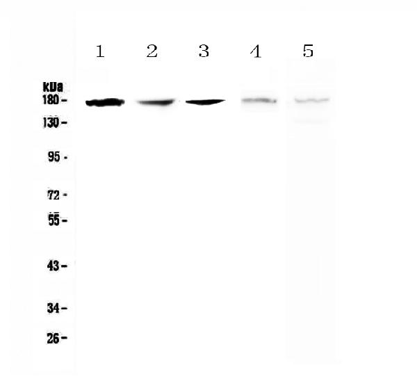
Figure 1. Western blot analysis of IRE1 using anti-IRE1 antibody (A00683-1). Electrophoresis was performed on a 5-20% SDS-PAGE gel at 70V (Stacking gel) / 90V (Resolving gel) for 2-3 hours. The sample well of each lane was loaded with 50ug of sample under reducing conditions. Lane 1: human A549 whole cell lysates, Lane 2: human SK-OV-3 whole cell lysates, Lane 3: human PANC-1 whole cell lysates, Lane 4: rat brain tissue lysates, Lane 5: mouse brain tissue lysates. After Electrophoresis, proteins were transferred to a Nitrocellulose membrane at 150mA for 50-90 minutes. Blocked the membrane with 5% Non-fat Milk/ TBS for 1.5 hour at RT. The membrane was incubated with rabbit anti-IRE1 antigen affinity purified polyclonal antibody (Catalog # A00683-1) at 0.5 microg/mL overnight at 4°C, then washed with TBS-0.1%Tween 3 times with 5 minutes each and probed with a goat anti-rabbit IgG-HRP secondary antibody at a dilution of 1:10000 for 1.5 hour at RT. The signal is developed using an Enhanced Chemiluminescent detection (ECL) kit (Catalog # EK1002) with Tanon 5200 system. A specific band was detected for IRE1 at approximately 170KD. The expected band size for IRE1 is at 110KD.
Figure 1. Western blot analysis of IRE1 using anti-IRE1 antibody (A00683-1). Electrophoresis was performed on a 5-20% SDS-PAGE gel at 70V (Stacking gel) / 90V (Resolving gel) for 2-3 hours. The sample well of each lane was loaded with 50ug of sample under reducing conditions. Lane 1: human A549 whole cell lysates, Lane 2: human SK-OV-3 whole cell lysates, Lane 3: human PANC-1 whole cell lysates, Lane 4: rat brain tissue lysates, Lane 5: mouse brain tissue lysates. After Electrophoresis, proteins were transferred to a Nitrocellulose membrane at 150mA for 50-90 minutes. Blocked the membrane with 5% Non-fat Milk/ TBS for 1.5 hour at RT. The membrane was incubated with rabbit anti-IRE1 antigen affinity purified polyclonal antibody (Catalog # A00683-1) at 0.5 microg/mL overnight at 4°C, then washed with TBS-0.1%Tween 3 times with 5 minutes each and probed with a goat anti-rabbit IgG-HRP secondary antibody at a dilution of 1:10000 for 1.5 hour at RT. The signal is developed using an Enhanced Chemiluminescent detection (ECL) kit (Catalog # EK1002) with Tanon 5200 system. A specific band was detected for IRE1 at approximately 170KD. The expected band size for IRE1 is at 110KD.
Anti-IRE1/ERN1 Antibody Picoband(r)
A00683-1-CARRIER-FREE
ApplicationsFlow Cytometry, ImmunoFluorescence, Western Blot, ELISA, ImmunoCytoChemistry
Product group Antibodies
ReactivityHuman, Mouse, Rat
TargetACTN4
Overview
- SupplierBoster Bio
- Product NameAnti-IRE1/ERN1 Antibody Picoband(r)
- Delivery Days Customer9
- ApplicationsFlow Cytometry, ImmunoFluorescence, Western Blot, ELISA, ImmunoCytoChemistry
- CertificationResearch Use Only
- ClonalityPolyclonal
- Concentration500 ug/ml
- Gene ID81
- Target nameACTN4
- Target descriptionactinin alpha 4
- Target synonymsACTININ-4, FSGS, FSGS1, alpha-actinin-4, focal segmental glomerulosclerosis 1, non-muscle alpha-actinin 4
- HostRabbit
- IsotypeIgG
- Protein IDO75460
- Protein NameSerine/threonine-protein kinase/endoribonuclease IRE1
- Scientific DescriptionBoster Bio Anti-IRE1/ERN1 Antibody Picoband® catalog # A00683-1. Tested in ELISA, Flow Cytometry, IF, ICC, WB applications. This antibody reacts with Human, Mouse, Rat. The brand Picoband indicates this is a premium antibody that guarantees superior quality, high affinity, and strong signals with minimal background in Western blot applications. Only our best-performing antibodies are designated as Picoband, ensuring unmatched performance.
- ReactivityHuman, Mouse, Rat
- Storage Instruction-20°C,2°C to 8°C
- UNSPSC12352203
Related products
Product group Antibodies
anti-alpha-Actinin-4, pAb (IG-701)AG-25T-0107
ApplicationsImmunoPrecipitation, Western Blot, ImmunoHistoChemistry
ReactivityHamster, Human, Mouse, Porcine, Rat
TargetACTN4
- SizePrice
Product group Antibodies

Anti-Alpha Actinin 4 [Ab01AA4]Ab02447-10.0
ApplicationsImmunoPrecipitation, ELISA, ImmunoHistoChemistry
ReactivityHuman
TargetACTN4
- SizePrice
Product group Antibodies
Anti-ACTN4 Antibody144-64273
ApplicationsImmunoFluorescence, Western Blot
ReactivityHuman, Mouse, Rat
TargetACTN4
- SizePrice
Product group Antibodies
Anti-ACTN4 AntibodyA42831
ApplicationsWestern Blot
ReactivityHuman, Mouse, Rat
- SizePrice
Product group Antibodies
alpha Actinin 4 Recombinant AntibodyBSM-60423R
ApplicationsImmunoFluorescence, Western Blot, ImmunoCytoChemistry, ImmunoHistoChemistry, ImmunoHistoChemistry Frozen, ImmunoHistoChemistry Paraffin
ReactivityHuman, Mouse
TargetACTN4
- SizePrice
Product group Antibodies
ACTN4 AntibodyCSB-PA00814A0RB
ApplicationsImmunoFluorescence, Western Blot, ELISA, ImmunoHistoChemistry
ReactivityHuman, Rat
TargetACTN4
- SizePrice
Product group Antibodies
Actn4 Polyclonal AntibodyCAC10364
ApplicationsImmunoFluorescence, Western Blot, ELISA, ImmunoHistoChemistry
ReactivityRat
TargetACTN4
- SizePrice
![alpha Actinin 4 antibody [C2C3], C-term detects ACTN4 protein by western blot analysis. A. 30 μg Neuro2A whole cell lysate/extract B. 30 μg GL261 whole cell lysate/extract C. 30 μg C8D30 whole cell lysate/extract D. 30 μg NIH-3T3 whole cell lysate/extract E. 30 μg BCL-1 whole cell lysate/extract F. 30 μg Raw264.7 whole cell lysate/extract G. 30 μg C2C12 whole cell lysate/extract 7.5% SDS-PAGE alpha Actinin 4 antibody [C2C3], C-term (GTX101669) dilution: 1:1000 The HRP-conjugated anti-rabbit IgG antibody (GTX213110-01) was used to detect the primary antibody.](https://www.genetex.com/upload/website/prouct_img/normal/GTX101669/GTX101669_40205_WB_M_w_23060100_117.webp)
Product group Antibodies
ApplicationsImmunoFluorescence, Western Blot, ImmunoCytoChemistry, ImmunoHistoChemistry, ImmunoHistoChemistry Paraffin
ReactivityHuman, Mouse, Rat
TargetACTN4
- SizePrice

Product group Antibodies
Anti-ACTN4 AntibodyHPA001873
ApplicationsWestern Blot, ImmunoCytoChemistry, ImmunoHistoChemistry
ReactivityHuman, Mouse, Rat
TargetACTN4
- SizePrice