

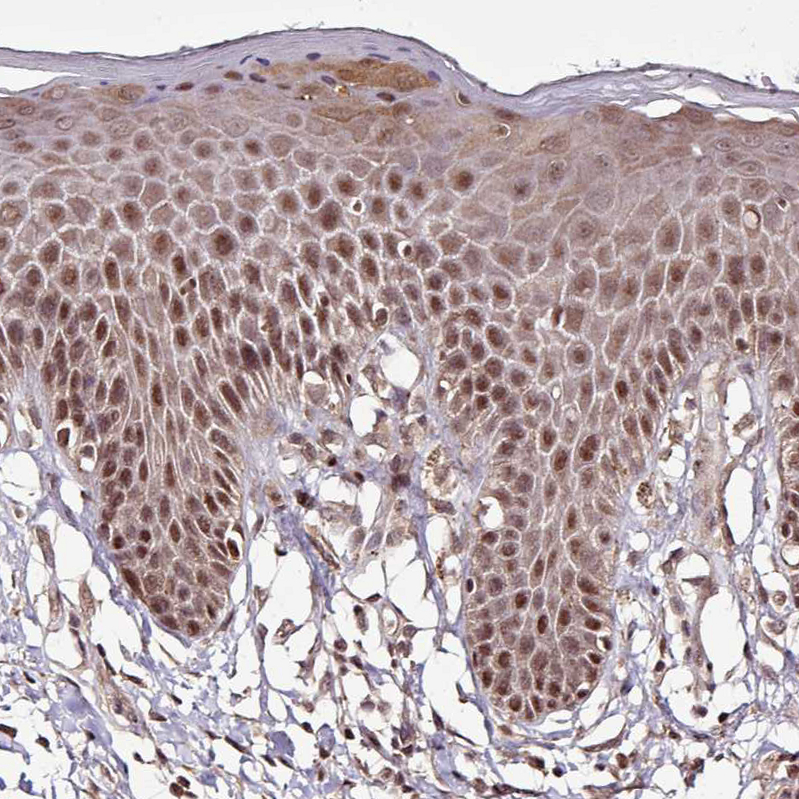
Immunohistochemical staining of human skin shows strong nuclear positivity in squamous epithelial cells.

Immunohistochemical staining of human skin shows strong nuclear positivity in squamous epithelial cells.
Anti-ITPKC Antibody
HPA053003
ApplicationsWestern Blot, ImmunoCytoChemistry, ImmunoHistoChemistry
Product group Antibodies
ReactivityHuman
TargetITPKC
Overview
- SupplierAtlas Antibodies
- Product NameAnti-ITPKC Antibody
- Delivery Days Customer4
- ApplicationsWestern Blot, ImmunoCytoChemistry, ImmunoHistoChemistry
- CertificationResearch Use Only
- ClonalityPolyclonal
- ConjugateUnconjugated
- Gene ID80271
- Target nameITPKC
- Target descriptioninositol-trisphosphate 3-kinase C
- Target synonymsIP3-3KC, IP3KC, inositol-trisphosphate 3-kinase C, IP3 3-kinase C, IP3K C, InsP 3 kinase C, inositol 1,4,5-trisphosphate 3-kinase C, insP 3-kinase C
- HostRabbit
- IsotypeIgG
- Protein IDQ96DU7
- Protein NameInositol-trisphosphate 3-kinase C
- Scientific DescriptionRecombinant Protein Epitope Signature Tag (PrEST) antigen sequence
- ReactivityHuman
- Storage Instruction-20°C,2°C to 8°C
- UNSPSC41116161
Datasheet
MSDS
Related products

Product group Antibodies
Anti-ITPKC Antibody Picoband(r)A07845-1-CARRIER-FREE
ApplicationsFlow Cytometry, Western Blot, ELISA
ReactivityHuman
TargetITPKC
- SizePrice
Product group Antibodies

ITPKC AntibodyLS-C830177
ApplicationsELISA, ImmunoHistoChemistry
ReactivityHuman
TargetITPKC
- SizePrice
Product group Antibodies
ITPKC AntibodyCSB-PA003041
ApplicationsWestern Blot, ELISA, ImmunoHistoChemistry
ReactivityHuman
TargetITPKC
- SizePrice
Product group Antibodies

Goat anti-IP3KCEB09030
ApplicationsWestern Blot, ELISA
ReactivityHuman
TargetITPKC
- SizePrice

Product group Antibodies
Anti-ITPKC AntibodyHPA050760
ApplicationsImmunoHistoChemistry
ReactivityHuman
TargetITPKC
- SizePrice
Product group Antibodies
IP3KC antibody [N3C2], InternalGTX104192
ApplicationsImmunoFluorescence, Western Blot, ImmunoCytoChemistry
ReactivityHuman
TargetITPKC
- SizePrice