


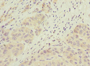
Immunohistochemical staining of human liver shows strong granular positivity in hepatocytes.

Immunohistochemical staining of human liver shows strong granular positivity in hepatocytes.
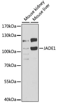
Anti-JADE1 Antibody
HPA020016
ApplicationsWestern Blot, ImmunoHistoChemistry
Product group Antibodies
ReactivityHuman, Mouse, Rat
TargetJADE1
Overview
- SupplierAtlas Antibodies
- Product NameAnti-JADE1 Antibody
- Delivery Days Customer4
- ApplicationsWestern Blot, ImmunoHistoChemistry
- CertificationResearch Use Only
- ClonalityPolyclonal
- ConjugateUnconjugated
- Gene ID79960
- Target nameJADE1
- Target descriptionjade family PHD finger 1
- Target synonymsPHF17, protein Jade-1, PHD finger protein 17, PHD protein Jade-1, gene for apoptosis and differentiation in epithelia, jade family PHD finger protein 1
- HostRabbit
- IsotypeIgG
- Protein IDQ6IE81
- Protein NameProtein Jade-1
- Scientific DescriptionRecombinant Protein Epitope Signature Tag (PrEST) antigen sequence
- ReactivityHuman, Mouse, Rat
- Storage Instruction-20°C,2°C to 8°C
- UNSPSC41116161
Datasheet
MSDS
Related products

Product group Antibodies
Anti-JADE1 Antibody Picoband(r)A07611-1-CARRIER-FREE
ApplicationsFlow Cytometry, ImmunoFluorescence, Western Blot, ELISA, ImmunoCytoChemistry
ReactivityHuman
TargetJADE1
- SizePrice
Product group Antibodies
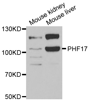
Anti-PHF17 AntibodyA31309
ApplicationsWestern Blot, ImmunoHistoChemistry
ReactivityHuman, Mouse, Rat
- SizePrice
Product group Antibodies

JADE1 / PHF17 AntibodyLS-C747674
ApplicationsImmunoFluorescence, ImmunoHistoChemistry
ReactivityHuman, Mouse
TargetJADE1
- SizePrice
Product group Antibodies
JADE1 AntibodyCSB-PA743563ESR1HU
ApplicationsELISA, ImmunoHistoChemistry
ReactivityHuman
TargetJADE1
- SizePrice
Product group Antibodies
PHF17 antibodyGTX32786
ApplicationsImmunoFluorescence, Western Blot, ImmunoCytoChemistry
ReactivityHuman, Mouse
TargetJADE1
- SizePrice