

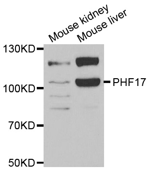
Anti-PHF17 Antibody
A31309
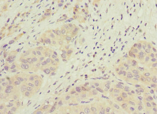
ApplicationsWestern Blot, ImmunoHistoChemistry
Product group Antibodies
ReactivityHuman, Mouse, Rat
Overview
- SupplierAntibodies.com
- Product NameAnti-PHF17 Antibody
- Delivery Days Customer7
- ApplicationsWestern Blot, ImmunoHistoChemistry
- CertificationResearch Use Only
- ClonalityPolyclonal
- ConjugateUnconjugated
- Estimated Purity>95%
- HostRabbit
- Scientific DescriptionRabbit polyclonal antibody to PHF17
- ReactivityHuman, Mouse, Rat
- UNSPSC12352203
Related products

Product group Antibodies
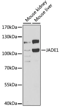
Anti-JADE1 Antibody Picoband(r)A07611-1-CARRIER-FREE
ApplicationsFlow Cytometry, ImmunoFluorescence, Western Blot, ELISA, ImmunoCytoChemistry
ReactivityHuman
TargetJADE1
- SizePrice
Product group Antibodies

JADE1 / PHF17 AntibodyLS-C747674
ApplicationsImmunoFluorescence, ImmunoHistoChemistry
ReactivityHuman, Mouse
TargetJADE1
- SizePrice

Product group Antibodies
Anti-JADE1 AntibodyHPA020016
ApplicationsWestern Blot, ImmunoHistoChemistry
ReactivityHuman, Mouse, Rat
TargetJADE1
- SizePrice
Product group Antibodies
JADE1 AntibodyCSB-PA743563ESR1HU
ApplicationsELISA, ImmunoHistoChemistry
ReactivityHuman
TargetJADE1
- SizePrice
Product group Antibodies
PHF17 antibodyGTX32786
ApplicationsImmunoFluorescence, Western Blot, ImmunoCytoChemistry
ReactivityHuman, Mouse
TargetJADE1
- SizePrice