


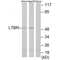
Figure 1. Western blot analysis of LTBR using anti-LTBR antibody (A00942-2). Electrophoresis was performed on a 5-20% SDS-PAGE gel at 70V (Stacking gel) / 90V (Resolving gel) for 2-3 hours. The sample well of each lane was loaded with 30 ug of sample under reducing conditions. Lane 1: human Hela whole cell lysates, Lane 2: human Hut-78 whole cell lysates, Lane 3: human K562 whole cell lysates, Lane 4: human HEL whole cell lysates. After electrophoresis, proteins were transferred to a nitrocellulose membrane at 150 mA for 50-90 minutes. Blocked the membrane with 5% non-fat milk/TBS for 1.5 hour at RT. The membrane was incubated with rabbit anti-LTBR antigen affinity purified polyclonal antibody (Catalog # A00942-2) at 0.5 microg/mL overnight at 4°C, then washed with TBS-0.1%Tween 3 times with 5 minutes each and probed with a goat anti-rabbit IgG-HRP secondary antibody at a dilution of 1:5000 for 1.5 hour at RT. The signal is developed using an Enhanced Chemiluminescent detection (ECL) kit (Catalog # EK1002) with Tanon 5200 system. A specific band was detected for LTBR at approximately 55-60 kDa. The expected band size for LTBR is at 47 kDa.
Figure 1. Western blot analysis of LTBR using anti-LTBR antibody (A00942-2). Electrophoresis was performed on a 5-20% SDS-PAGE gel at 70V (Stacking gel) / 90V (Resolving gel) for 2-3 hours. The sample well of each lane was loaded with 30 ug of sample under reducing conditions. Lane 1: human Hela whole cell lysates, Lane 2: human Hut-78 whole cell lysates, Lane 3: human K562 whole cell lysates, Lane 4: human HEL whole cell lysates. After electrophoresis, proteins were transferred to a nitrocellulose membrane at 150 mA for 50-90 minutes. Blocked the membrane with 5% non-fat milk/TBS for 1.5 hour at RT. The membrane was incubated with rabbit anti-LTBR antigen affinity purified polyclonal antibody (Catalog # A00942-2) at 0.5 microg/mL overnight at 4°C, then washed with TBS-0.1%Tween 3 times with 5 minutes each and probed with a goat anti-rabbit IgG-HRP secondary antibody at a dilution of 1:5000 for 1.5 hour at RT. The signal is developed using an Enhanced Chemiluminescent detection (ECL) kit (Catalog # EK1002) with Tanon 5200 system. A specific band was detected for LTBR at approximately 55-60 kDa. The expected band size for LTBR is at 47 kDa.
Anti-LTBR Antibody Picoband(r)
A00942-2-CARRIER-FREE
ApplicationsWestern Blot, ELISA
Product group Antibodies
ReactivityHuman
TargetLTBR
Overview
- SupplierBoster Bio
- Product NameAnti-LTBR Antibody Picoband(r)
- Delivery Days Customer9
- ApplicationsWestern Blot, ELISA
- CertificationResearch Use Only
- ClonalityPolyclonal
- Concentration500 ug/ml
- Gene ID4055
- Target nameLTBR
- Target descriptionlymphotoxin beta receptor
- Target synonymsD12S370, LT-BETA-R, TNF-R-III, TNFCR, TNFR-RP, TNFR2-RP, TNFR3, TNFRSF3, tumor necrosis factor receptor superfamily member 3, lymphotoxin B receptor, lymphotoxin beta receptor (TNFR superfamily, member 3), tumor necrosis factor C receptor, tumor necrosis factor receptor 2-related protein, tumor necrosis factor receptor type III
- HostRabbit
- Protein IDP36941
- Protein NameTumor necrosis factor receptor superfamily member 3
- Scientific DescriptionBoster Bio Anti-LTBR Antibody Picoband® catalog # A00942-2. Tested in WB, ELISA applications. This antibody reacts with Human. The brand Picoband indicates this is a premium antibody that guarantees superior quality, high affinity, and strong signals with minimal background in Western blot applications. Only our best-performing antibodies are designated as Picoband, ensuring unmatched performance.
- ReactivityHuman
- Storage Instruction-20°C,2°C to 8°C
- UNSPSC12352203
Related products
Product group Antibodies
Anti-LTBR AntibodyA39936
ApplicationsImmunoFluorescence, Western Blot
ReactivityHuman, Mouse
- SizePrice
Product group Antibodies
Anti-LTBR Antibody144-65892
ApplicationsWestern Blot
ReactivityHuman, Mouse
TargetLTBR
- SizePrice
Product group Antibodies
LTBR Polyclonal AntibodyBS-6917R
ApplicationsImmunoFluorescence, Western Blot, ELISA, ImmunoCytoChemistry, ImmunoHistoChemistry, ImmunoHistoChemistry Frozen, ImmunoHistoChemistry Paraffin
ReactivityBovine, Human, Mouse, Porcine, Rat
TargetLTBR
- SizePrice
Product group Antibodies
LTBR AntibodyCSB-PA003174
ApplicationsImmunoFluorescence, Western Blot, ELISA
ReactivityHuman, Mouse
TargetLTBR
- SizePrice
Product group Antibodies
ApplicationsWestern Blot, ImmunoHistoChemistry
TargetLTBR
- SizePrice

Product group Antibodies
Anti-LTBR AntibodyHPA061617
ApplicationsImmunoCytoChemistry
ReactivityHuman
TargetLTBR
- SizePrice
Product group Antibodies
LTBR antibodyGTX10493
ApplicationsFlow Cytometry, Western Blot
ReactivityHuman
TargetLTBR
- SizePrice
Product group Antibodies

LTBR AntibodyLS-C747223
ApplicationsWestern Blot
ReactivityHuman, Mouse
TargetLTBR
- SizePrice
Product group Antibodies
Anti-LTBR AntibodyCAB5351
ApplicationsImmunoFluorescence, Western Blot, ELISA, ImmunoCytoChemistry, ImmunoHistoChemistry, ImmunoHistoChemistry Paraffin
ReactivityHuman
TargetLTBR
- SizePrice