


Figure 2. Flow Cytometry analysis of PC-3 cells using anti-MAG antibody (A03019). Overlay histogram showing PC-3 cells stained with A03019 (Blue line). To facilitate intracellular staining, cells were fixed with 4% paraformaldehyde and permeabilized with permeabilization buffer. The cells were blocked with 10% normal goat serum. And then incubated with rabbit anti-MAG Antibody (A03019, 1microg/1x106 cells) for 30 min at 20°C. DyLight®488 conjugated goat anti-rabbit IgG (BA1127, 5-10microg/1x106 cells) was used as secondary antibody for 30 minutes at 20°C. Isotype control antibody (Green line) was rabbit IgG (1microg/1x106) used under the same conditions. Unlabelled sample without incubation with primary antibody and secondary antibody (Red line) was used as a blank control.
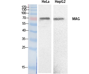
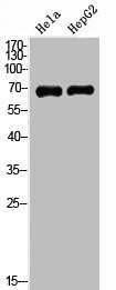
. Electrophoresis was performed on a 5-20% SDS-PAGE gel at 70V (Stacking gel) / 90V (Resolving gel) for 2-3 hours. The sample well of each lane was loaded with 30 ug of sample under reducing conditions. Lane 1: rat brain tissue lysates, Lane 2: mouse brain tissue lysates. After electrophoresis, proteins were transferred to a nitrocellulose membrane at 150 mA for 50-90 minutes. Blocked the membrane with 5% non-fat milk/TBS for 1.5 hour at RT. The membrane was incubated with rabbit anti-MAG antigen affinity purified polyclonal antibody (Catalog # A03019) at 0.5 microg/mL overnight at 4°C, then washed with TBS-0.1%Tween 3 times with 5 minutes each and probed with a goat anti-rabbit IgG-HRP secondary antibody at a dilution of 1:5000 for 1.5 hour at RT. The signal is developed using an Enhanced Chemiluminescent detection (ECL) kit (Catalog # EK1002) with Tanon 5200 system. A specific band was detected for MAG at approximately 100 kDa. The expected band size for MAG is at 69 kDa.")
. Overlay histogram showing K562 cells stained with A03019 (Blue line). The cells were fixed with 4% paraformaldehyde and blocked with 10% normal goat serum. And then incubated with rabbit anti-MAG Antibody (A03019, 1 microg/1x106 cells) for 30 min at 20°C. DyLight®488 conjugated goat anti-rabbit IgG (BA1127, 5-10 microg/1x106 cells) was used as secondary antibody for 30 minutes at 20°C. Isotype control antibody (Green line) was rabbit IgG (1 microg/1x106) used under the same conditions. Unlabelled sample without incubation with primary antibody and secondary antibody (Red line) was used as a blank control.")
Figure 2. Flow Cytometry analysis of PC-3 cells using anti-MAG antibody (A03019). Overlay histogram showing PC-3 cells stained with A03019 (Blue line). To facilitate intracellular staining, cells were fixed with 4% paraformaldehyde and permeabilized with permeabilization buffer. The cells were blocked with 10% normal goat serum. And then incubated with rabbit anti-MAG Antibody (A03019, 1microg/1x106 cells) for 30 min at 20°C. DyLight®488 conjugated goat anti-rabbit IgG (BA1127, 5-10microg/1x106 cells) was used as secondary antibody for 30 minutes at 20°C. Isotype control antibody (Green line) was rabbit IgG (1microg/1x106) used under the same conditions. Unlabelled sample without incubation with primary antibody and secondary antibody (Red line) was used as a blank control.
Anti-MAG Antibody Picoband(r)
A03019-CARRIER-FREE
ApplicationsFlow Cytometry, Western Blot, ELISA
Product group Antibodies
ReactivityHuman, Mouse, Rat
TargetMAG
Overview
- SupplierBoster Bio
- Product NameAnti-MAG Antibody Picoband(r)
- Delivery Days Customer9
- Application Supplier NoteTested Species: In-house tested species with positive results. Other applications have not been tested. Optimal dilutions should be determined by end users.
- ApplicationsFlow Cytometry, Western Blot, ELISA
- CertificationResearch Use Only
- ClonalityPolyclonal
- Concentration500 ug/ml
- Gene ID4099
- Target nameMAG
- Target descriptionmyelin associated glycoprotein
- Target synonymsGMA, S-MAG, SIGLEC-4A, SIGLEC4, SIGLEC4A, SPG75, myelin-associated glycoprotein, sialic acid binding Ig-like lectin 4A, sialic acid-binding immunoglobulin-like lectin 4A
- HostRabbit
- IsotypeIgG
- Protein IDP20916
- Protein NameMyelin-associated glycoprotein
- Scientific DescriptionBoster Bio Anti-MAG Antibody Picoband® catalog # A03019. Tested in ELISA, Flow Cytometry, WB applications. This antibody reacts with Human, Mouse, Rat. The brand Picoband indicates this is a premium antibody that guarantees superior quality, high affinity, and strong signals with minimal background in Western blot applications. Only our best-performing antibodies are designated as Picoband, ensuring unmatched performance.
- ReactivityHuman, Mouse, Rat
- Storage Instruction-20°C,2°C to 8°C
- UNSPSC12352203
Related products
Product group Antibodies
Anti-MAG AntibodyA98483
ApplicationsWestern Blot, ELISA
ReactivityHuman, Mouse, Rat
- SizePrice
Product group Antibodies
Anti-MAG Antibody144-07186
ApplicationsWestern Blot
ReactivityHuman, Mouse, Rat
TargetMAG
- SizePrice
Product group Antibodies
References
MAG Polyclonal AntibodyBS-0257R
ApplicationsImmunoFluorescence, Western Blot, ELISA, ImmunoCytoChemistry, ImmunoHistoChemistry, ImmunoHistoChemistry Frozen, ImmunoHistoChemistry Paraffin
ReactivityHuman, Mouse, Rat
TargetMAG
- SizePrice
Product group Antibodies
MAG AntibodyCSB-PA006022
ApplicationsWestern Blot, ELISA
ReactivityHuman, Mouse, Rat
TargetMAG
- SizePrice
Product group Antibodies
MAG Polyclonal AntibodyCAC13105
ApplicationsImmunoFluorescence, Western Blot, ELISA, ImmunoHistoChemistry
ReactivityMouse, Rat
TargetMAG
- SizePrice
Product group Antibodies

MAG AntibodyLS-C400865
ApplicationsWestern Blot, ELISA, ImmunoHistoChemistry
ReactivityHuman, Mouse, Rat
TargetMAG
- SizePrice

Product group Antibodies
Anti-MAG AntibodyHPA012499
ApplicationsImmunoHistoChemistry
ReactivityHuman
TargetMAG
- SizePrice
Product group Antibodies
MAG antibodyGTX114542
ApplicationsWestern Blot, ImmunoHistoChemistry, ImmunoHistoChemistry Frozen, ImmunoHistoChemistry Paraffin
ReactivityHuman, Mouse, Rat
TargetMAG
- SizePrice
Product group Antibodies
Anti-MAG AntibodyCAB7186
ApplicationsWestern Blot, ELISA
ReactivityHuman
TargetMAG
- SizePrice