

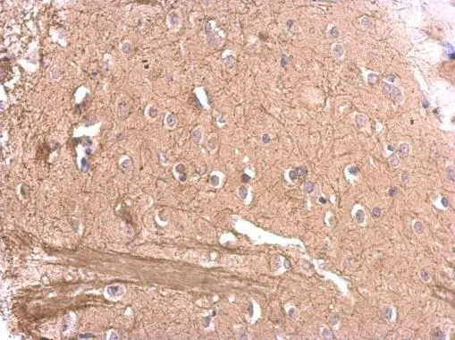
MAG antibody detects MAG protein at on rat fore brain by immunohistochemical analysis. Sample: Paraffin-embedded rat fore brain. MAG antibody (GTX114542) dilution: 1:500.
Antigen Retrieval: Trilogy? (EDTA based, pH 8.0) buffer, 15min
![MAG antibody detects MAG protein expression by immunohistochemical analysis. Sample: Frozen-sectioned adult mouse cerebellum. Green: MAG protein stained by MAG antibody (GTX114542) diluted at 1:250. Red: beta Tubulin 3/ TUJ1, stained by beta Tubulin 3/ TUJ1 antibody [GT11710] (GTX631836) diluted at 1:500. Blue: Fluoroshield with DAPI (GTX30920).](https://www.genetex.com/upload/website/prouct_img/normal/GTX114542/GTX114542_40198_20170608_IHC-P_M_w_23060518_581.webp "MAG antibody detects MAG protein expression by immunohistochemical analysis. Sample: Frozen-sectioned adult mouse cerebellum. Green: MAG protein stained by MAG antibody (GTX114542) diluted at 1:250. Red: beta Tubulin 3/ TUJ1, stained by beta Tubulin 3/ TUJ1 antibody [GT11710] (GTX631836) diluted at 1:500. Blue: Fluoroshield with DAPI (GTX30920).")
 was separated by 7.5% SDS-PAGE, and the membrane was blotted with MAG antibody (GTX114542) diluted at 1:1000.")
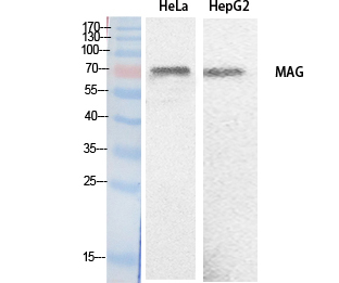
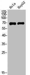
 were separated by 7.5% SDS-PAGE, and the membrane was blotted with MAG antibody (GTX114542) diluted at 1:1000. The HRP-conjugated anti-rabbit IgG antibody (GTX213110-01) was used to detect the primary antibody.")
 were separated by 7.5% SDS-PAGE, and the membrane was blotted with MAG antibody (GTX114542) diluted at 1:1000. The HRP-conjugated anti-rabbit IgG antibody (GTX213110-01) was used to detect the primary antibody.")
MAG antibody detects MAG protein at on rat fore brain by immunohistochemical analysis. Sample: Paraffin-embedded rat fore brain. MAG antibody (GTX114542) dilution: 1:500.
Antigen Retrieval: Trilogy? (EDTA based, pH 8.0) buffer, 15min
MAG antibody
GTX114542
ApplicationsWestern Blot, ImmunoHistoChemistry, ImmunoHistoChemistry Frozen, ImmunoHistoChemistry Paraffin
Product group Antibodies
ReactivityHuman, Mouse, Rat
TargetMAG
Overview
- SupplierGeneTex
- Product NameMAG antibody
- Delivery Days Customer9
- Application Supplier NoteWB: 1:500-1:3000. IHC-P: 1:100-1:1000. IHC-Fr: 1:100-1:1000. *Optimal dilutions/concentrations should be determined by the researcher.Not tested in other applications.
- ApplicationsWestern Blot, ImmunoHistoChemistry, ImmunoHistoChemistry Frozen, ImmunoHistoChemistry Paraffin
- CertificationResearch Use Only
- ClonalityPolyclonal
- Concentration0.61 mg/ml
- ConjugateUnconjugated
- Gene ID4099
- Target nameMAG
- Target descriptionmyelin associated glycoprotein
- Target synonymsGMA, S-MAG, SIGLEC-4A, SIGLEC4, SIGLEC4A, SPG75, myelin-associated glycoprotein, sialic acid binding Ig-like lectin 4A, sialic acid-binding immunoglobulin-like lectin 4A
- HostRabbit
- IsotypeIgG
- Protein IDP20916
- Protein NameMyelin-associated glycoprotein
- Scientific DescriptionThe protein encoded by this gene is a type I membrane protein and member of the immunoglobulin superfamily. It is thought to be involved in the process of myelination. It is a lectin that binds to sialylated glycoconjugates and mediates certain myelin-neuron cell-cell interactions. Two alternatively spliced transcripts encoding different isoforms have been described for this gene. [provided by RefSeq]
- ReactivityHuman, Mouse, Rat
- Storage Instruction-20°C or -80°C,2°C to 8°C
- UNSPSC41116161
Datasheet
Related products
Product group Antibodies
Anti-MAG AntibodyA98483
ApplicationsWestern Blot, ELISA
ReactivityHuman, Mouse, Rat
- SizePrice

Product group Antibodies
Anti-MAG Antibody Picoband(r)A03019-CARRIER-FREE
ApplicationsFlow Cytometry, Western Blot, ELISA
ReactivityHuman, Mouse, Rat
TargetMAG
- SizePrice
Product group Antibodies
Anti-MAG Antibody144-07186
ApplicationsWestern Blot
ReactivityHuman, Mouse, Rat
TargetMAG
- SizePrice
Product group Antibodies
References
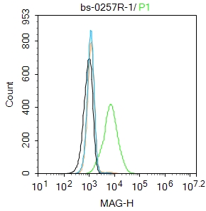
MAG Polyclonal AntibodyBS-0257R
ApplicationsImmunoFluorescence, Western Blot, ELISA, ImmunoCytoChemistry, ImmunoHistoChemistry, ImmunoHistoChemistry Frozen, ImmunoHistoChemistry Paraffin
ReactivityHuman, Mouse, Rat
TargetMAG
- SizePrice
Product group Antibodies
MAG AntibodyCSB-PA006022
ApplicationsWestern Blot, ELISA
ReactivityHuman, Mouse, Rat
TargetMAG
- SizePrice
Product group Antibodies
MAG Polyclonal AntibodyCAC13105
ApplicationsImmunoFluorescence, Western Blot, ELISA, ImmunoHistoChemistry
ReactivityMouse, Rat
TargetMAG
- SizePrice
Product group Antibodies

MAG AntibodyLS-C400865
ApplicationsWestern Blot, ELISA, ImmunoHistoChemistry
ReactivityHuman, Mouse, Rat
TargetMAG
- SizePrice

Product group Antibodies
Anti-MAG AntibodyHPA012499
ApplicationsImmunoHistoChemistry
ReactivityHuman
TargetMAG
- SizePrice
![Boiled and unboiled various tissue extracts (50 μg) were separated by 7.5% SDS-PAGE, and the membrane was blotted with MAG antibody [HL3467] (GTX641352) diluted at 1:1000. The HRP-conjugated anti-rabbit IgG antibody (GTX213110-01) was used to detect the primary antibody.](https://www.genetex.com/upload/website/prouct_img/normal/GTX641352/GTX641352_T-45600_20241122_WB_M_tissue_24112622_869.webp)
Product group Antibodies
MAG antibody [HL3467]GTX641352
ApplicationsWestern Blot, ImmunoHistoChemistry, ImmunoHistoChemistry Paraffin
ReactivityHuman, Mouse, Rat
TargetMAG
- SizePrice
![Boiled and unboiled various tissue extracts (50 μg) were separated by 7.5% SDS-PAGE, and the membrane was blotted with MAG antibody [HL3469] (GTX641354) diluted at 1:1000. The HRP-conjugated anti-rabbit IgG antibody (GTX213110-01) was used to detect the primary antibody.](https://www.genetex.com/upload/website/prouct_img/normal/GTX641354/GTX641354_T-45600_20241122_WB_M_tissue_24112622_860.webp)
Product group Antibodies
MAG antibody [HL3469]GTX641354
ApplicationsWestern Blot, ImmunoHistoChemistry, ImmunoHistoChemistry Frozen, ImmunoHistoChemistry Paraffin
ReactivityHuman, Mouse, Rat
TargetMAG
- SizePrice