Bio-Connect
Anti-MFN1 Antibody

Anti-MFN1 Antibody

A46714
Antibodies.com
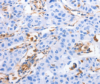
ApplicationsImmunoHistoChemistry
Product group Antibodies
Sign in to order and to see your custom pricing.
Large volume orders?
Order with a bulk request

Overview

  • Supplier
    Antibodies.com
  • Product Name
    Anti-MFN1 Antibody
  • Delivery Days Customer
    7
  • Applications
    ImmunoHistoChemistry
  • Certification
    Research Use Only
  • Clonality
    Polyclonal
  • Concentration
    2.2 mg/ml
  • Conjugate
    Unconjugated
  • Host
    Rabbit
  • Scientific Description
    Rabbit polyclonal antibody to MFN1
  • UNSPSC
    12352203

Related products

Product group Antibodies
Everest Biotech
ApplicationsELISA, ImmunoHistoChemistry
TargetMFN1
  • SizePrice
Western blot All lanes: MFN1 antibody at 3.78microg/ml Lane 1: Human placenta tissue Lane 2: Mouse kidney tissue Lane 3: NIH/3T3 whole cell lysate Lane 4: Jurkat whole cell lysate Lane 5: Hela whole cell lysate Lane 6: K562 whole cell lysate Secondary Goat polyclonal to rabbit IgG at 1/10000 dilution Predicted band size: 85, 42, 72 kDa Observed band size: 85 kDa
Product group Antibodies
Cusabio
MFN1 AntibodyCSB-PA812879ESR1HU
ApplicationsWestern Blot, ELISA, ImmunoHistoChemistry
TargetMFN1
  • SizePrice
Product group Antibodies
Assay Genie
TargetMFN1
  • SizePrice
RSC96 probed with Mfn1 Polyclonal Antibody, Unconjugated (bs-0557R) at 1:100 for 30 minutes followed by incubation with a conjugated secondary (PE Conjugated) (green) for 30 minutes compared to control cells (blue), secondary only (light blue) and isotype control (orange).
Product group Antibodies
References
Bioss Antibodies
ApplicationsFlow Cytometry, Western Blot, ELISA, ImmunoHistoChemistry, ImmunoHistoChemistry Paraffin
TargetMFN1
  • SizePrice
Product group Antibodies
Biomatik
ApplicationsWestern Blot, ELISA, ImmunoHistoChemistry
TargetMFN1
  • SizePrice
WB analysis of A431 cell lysate using GTX17218 MFN1 antibody. Working concentration : 1 microg/ml
Product group Antibodies
GeneTex
ApplicationsImmunoFluorescence, Western Blot, ELISA, ImmunoCytoChemistry
TargetMFN1
  • SizePrice
Immunofluorescent staining of human cell line U-2 OS shows localization to mitochondria.
Product group Antibodies
Atlas Antibodies
ApplicationsImmunoCytoChemistry
TargetMFN1
  • SizePrice
Product group Antibodies
RayBiotech
ApplicationsImmunoFluorescence, Western Blot
TargetMFN1
  • SizePrice
Product group Antibodies
LifeSpan BioSciences
MFN1 AntibodyLS-C400878
ApplicationsELISA, ImmunoHistoChemistry
TargetMFN1
  • SizePrice
Figure 1. Western blot analysis of Mitofusin 1 using anti-Mitofusin 1 antibody (PB9264). Electrophoresis was performed on a 5-20% SDS-PAGE gel at 70V (Stacking gel) / 90V (Resolving gel) for 2-3 hours. The sample well of each lane was loaded with 50ug of sample under reducing conditions. Lane 1: rat kidney tissue lysates, Lane 2: rat heart tissue lysates, Lane 3: mouse kidney tissue lysates, Lane 4: mouse heart tissue lysates, Lane 5: human placenta tissue lysates. After Electrophoresis, proteins were transferred to a Nitrocellulose membrane at 150mA for 50-90 minutes. Blocked the membrane with 5% Non-fat Milk/ TBS for 1.5 hour at RT. The membrane was incubated with rabbit anti-Mitofusin 1 antigen affinity purified polyclonal antibody (Catalog # PB9264) at 0.5 microg/mL overnight at 4°C, then washed with TBS-0.1%Tween 3 times with 5 minutes each and probed with a goat anti-rabbit IgG-HRP secondary antibody at a dilution of 1:10000 for 1.5 hour at RT. The signal is developed using an Enhanced Chemiluminescent detection (ECL) kit (Catalog # EK1002) with Tanon 5200 system. A specific band was detected for Mitofusin 1 at approximately 84KD. The expected band size for Mitofusin 1 is at 84KD.
Product group Antibodies
Boster Bio
ApplicationsWestern Blot
TargetMFN1
  • SizePrice