

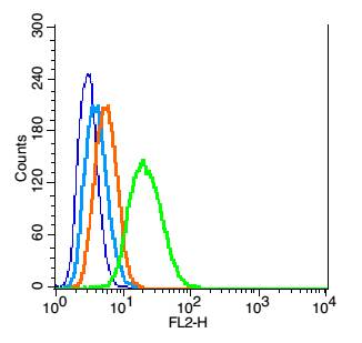
RSC96 probed with Mfn1 Polyclonal Antibody, Unconjugated (bs-0557R) at 1:100 for 30 minutes followed by incubation with a conjugated secondary (PE Conjugated) (green) for 30 minutes compared to control cells (blue), secondary only (light blue) and isotype control (orange).
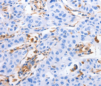
 for 15min; Block endogenous peroxidase by 3% hydrogen peroxide for 30 minutes; Blocking buffer (normal goat serum) at 37°C for 20min; Antibody incubation with Mitofusin 1 Polyclonal Antibody, Unconjugated (bs-0557R) at 1:400 overnight at 4°C, followed by a conjugated secondary for 20 minutes and DAB staining.")
 for 15min; Block endogenous peroxidase by 3% hydrogen peroxide for 30 minutes; Blocking buffer (normal goat serum) at 37°C for 20min; Antibody incubation with Mitofusin 1 Polyclonal Antibody, Unconjugated (bs-0557R) at 1:400 overnight at 4°C, followed by a conjugated secondary for 20 minutes and DAB staining.")
RSC96 probed with Mfn1 Polyclonal Antibody, Unconjugated (bs-0557R) at 1:100 for 30 minutes followed by incubation with a conjugated secondary (PE Conjugated) (green) for 30 minutes compared to control cells (blue), secondary only (light blue) and isotype control (orange).
Mitofusin 1 Polyclonal Antibody
BS-0557R
ApplicationsFlow Cytometry, Western Blot, ELISA, ImmunoHistoChemistry, ImmunoHistoChemistry Paraffin
Product group Antibodies
ReactivityEquine, Human, Mouse, Rabbit, Rat
TargetMFN1
Overview
- SupplierBioss
- Product NameMitofusin 1 Polyclonal Antibody
- Delivery Days Customer16
- ApplicationsFlow Cytometry, Western Blot, ELISA, ImmunoHistoChemistry, ImmunoHistoChemistry Paraffin
- Applications SupplierWB(1:300-5000), ELISA(1:500-1000), FCM(1:20-100), IHC-P(1:200-400)
- CertificationResearch Use Only
- ClonalityPolyclonal
- Concentration1 ug/ul
- ConjugateUnconjugated
- Gene ID55669
- Target nameMFN1
- Target descriptionmitofusin 1
- Target synonymshfzo1, hfzo2, mitofusin-1, fzo homolog, mitochondrial transmembrane GTPase FZO-2, mitochondrial transmembrane GTPase Fzo-1, putative transmembrane GTPase, transmembrane GTPase MFN1
- HostRabbit
- IsotypeIgG
- Protein IDQ8IWA4
- Protein NameMitofusin-1
- ReactivityEquine, Human, Mouse, Rabbit, Rat
- Storage Instruction-20°C
- UNSPSC41116161
References
- Hydrogen sulfide-induced oxidative stress leads to excessive mitochondrial fission to activate apoptosis in broiler myocardia. Wang S et al., 2019 Nov 15, Ecotoxicol Environ SafRead this paper
- PM2.5, SO2 and NO2 co-exposure impairs neurobehavior and induces mitochondrial injuries in the mouse brain. Ku T et al., 2016 Nov, ChemosphereRead this paper
Datasheet
Related products
Product group Antibodies
Anti-MFN1 AntibodyA46714
ApplicationsImmunoHistoChemistry
ReactivityHuman
- SizePrice

Product group Antibodies
Anti-MFN1 AntibodyHPA059230
ApplicationsImmunoCytoChemistry
ReactivityHuman
TargetMFN1
- SizePrice
Product group Antibodies

Goat anti-Mitofusin 1EB08139
ApplicationsELISA, ImmunoHistoChemistry
ReactivityHuman, Mouse, Rat
TargetMFN1
- SizePrice
Product group Antibodies
MFN1 AntibodyCSB-PA812879ESR1HU
ApplicationsWestern Blot, ELISA, ImmunoHistoChemistry
ReactivityHuman, Mouse
TargetMFN1
- SizePrice
Product group Antibodies

MFN1 AntibodyLS-C400878
ApplicationsELISA, ImmunoHistoChemistry
ReactivityHuman, Mouse, Rat
TargetMFN1
- SizePrice
Product group Antibodies
Mfn1 Polyclonal AntibodyCAC10824
ApplicationsWestern Blot, ELISA, ImmunoHistoChemistry
ReactivityMouse
TargetMFN1
- SizePrice

Product group Antibodies
Anti-Mitofusin 1/MFN1 Antibody Picoband(r)PB9264-CARRIER-FREE
ApplicationsWestern Blot
ReactivityBovine, Human, Mouse, Rat
TargetMFN1
- SizePrice
Product group Antibodies
MFN1 antibodyGTX17218
ApplicationsImmunoFluorescence, Western Blot, ELISA, ImmunoCytoChemistry
ReactivityHuman, Mouse, Rat
TargetMFN1
- SizePrice
Product group Antibodies
Anti-MFN1 Antibody144-09880
ApplicationsImmunoFluorescence, Western Blot
ReactivityHuman, Mouse, Rat
TargetMFN1
- SizePrice