


Immunofluorescent staining of human cell line U-2 OS shows localization to mitochondria.
Immunofluorescent staining of human cell line U-2 OS shows localization to mitochondria.
Anti-MFN1 Antibody
HPA059230
ApplicationsImmunoCytoChemistry
Product group Antibodies
ReactivityHuman
TargetMFN1
Overview
- SupplierAtlas Antibodies
- Product NameAnti-MFN1 Antibody
- Delivery Days Customer4
- ApplicationsImmunoCytoChemistry
- CertificationResearch Use Only
- ClonalityPolyclonal
- ConjugateUnconjugated
- Gene ID55669
- Target nameMFN1
- Target descriptionmitofusin 1
- Target synonymshfzo1, hfzo2, mitofusin-1, fzo homolog, mitochondrial transmembrane GTPase FZO-2, mitochondrial transmembrane GTPase Fzo-1, putative transmembrane GTPase, transmembrane GTPase MFN1
- HostRabbit
- IsotypeIgG
- Scientific DescriptionRecombinant Protein Epitope Signature Tag (PrEST) antigen sequence
- ReactivityHuman
- Storage Instruction-20°C,2°C to 8°C
- UNSPSC41116161
Datasheet
MSDS
Related products
Product group Antibodies
Anti-MFN1 AntibodyA46714
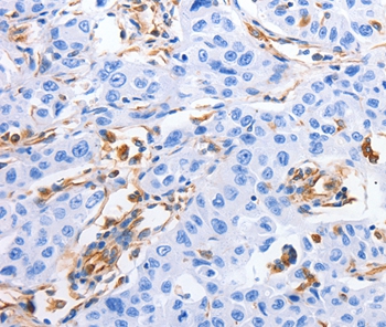
ApplicationsImmunoHistoChemistry
ReactivityHuman
- SizePrice
Product group Antibodies

Goat anti-Mitofusin 1EB08139
ApplicationsELISA, ImmunoHistoChemistry
ReactivityHuman, Mouse, Rat
TargetMFN1
- SizePrice
Product group Antibodies
MFN1 AntibodyCSB-PA812879ESR1HU
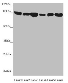
ApplicationsWestern Blot, ELISA, ImmunoHistoChemistry
ReactivityHuman, Mouse
TargetMFN1
- SizePrice
Product group Antibodies

MFN1 AntibodyLS-C400878
ApplicationsELISA, ImmunoHistoChemistry
ReactivityHuman, Mouse, Rat
TargetMFN1
- SizePrice
Product group Antibodies
Mfn1 Polyclonal AntibodyCAC10824
ApplicationsWestern Blot, ELISA, ImmunoHistoChemistry
ReactivityMouse
TargetMFN1
- SizePrice

Product group Antibodies
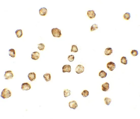
Anti-Mitofusin 1/MFN1 Antibody Picoband(r)PB9264-CARRIER-FREE
ApplicationsWestern Blot
ReactivityBovine, Human, Mouse, Rat
TargetMFN1
- SizePrice
Product group Antibodies
References
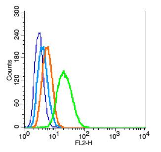
Mitofusin 1 Polyclonal AntibodyBS-0557R
ApplicationsFlow Cytometry, Western Blot, ELISA, ImmunoHistoChemistry, ImmunoHistoChemistry Paraffin
ReactivityEquine, Human, Mouse, Rabbit, Rat
TargetMFN1
- SizePrice
Product group Antibodies
MFN1 antibodyGTX17218
ApplicationsImmunoFluorescence, Western Blot, ELISA, ImmunoCytoChemistry
ReactivityHuman, Mouse, Rat
TargetMFN1
- SizePrice
Product group Antibodies
Anti-MFN1 Antibody144-09880
ApplicationsImmunoFluorescence, Western Blot
ReactivityHuman, Mouse, Rat
TargetMFN1
- SizePrice