


Immunofluorescent staining of human cell line HUVEC TERT2 shows localization to plasma membrane & cell junctions.
Immunofluorescent staining of human cell line HUVEC TERT2 shows localization to plasma membrane & cell junctions.
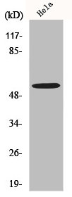
Anti-MLKL Antibody
HPA078638
ApplicationsImmunoCytoChemistry
Product group Antibodies
ReactivityHuman
TargetMLKL
Overview
- SupplierAtlas Antibodies
- Product NameAnti-MLKL Antibody
- Delivery Days Customer4
- ApplicationsImmunoCytoChemistry
- CertificationResearch Use Only
- ClonalityPolyclonal
- ConjugateUnconjugated
- Gene ID197259
- Target nameMLKL
- Target descriptionmixed lineage kinase domain like pseudokinase
- Target synonymshMLKL, mixed lineage kinase domain-like protein
- HostRabbit
- IsotypeIgG
- Protein IDQ8NB16
- Protein NameMixed lineage kinase domain-like protein
- Scientific DescriptionRecombinant Protein Epitope Signature Tag (PrEST) antigen sequence
- ReactivityHuman
- Storage Instruction-20°C,2°C to 8°C
- UNSPSC41116161
Datasheet
MSDS
Related products
Product group Antibodies
MLKL AntibodyCSB-PA003250
ApplicationsWestern Blot, ELISA
ReactivityHuman
TargetMLKL
- SizePrice

Product group Antibodies
Anti-MLKL Antibody Picoband(r)A00535-3-CARRIER-FREE
ApplicationsFlow Cytometry, ImmunoFluorescence, Western Blot, ELISA, ImmunoCytoChemistry, ImmunoHistoChemistry
ReactivityHuman, Mouse, Rat
TargetMLKL
- SizePrice
Product group Antibodies

MLKL Antibody (clone 8H7)LS-C768174
ApplicationsImmunoHistoChemistry, ImmunoHistoChemistry Paraffin
ReactivityHuman
TargetMLKL
- SizePrice
Product group Antibodies
Mlkl Polyclonal AntibodyCAC10516
ApplicationsELISA, ImmunoHistoChemistry
TargetMLKL
- SizePrice

Product group Antibodies
MLKL Recombinant AntibodyBSM-52256R
ApplicationsWestern Blot
ReactivityHuman
TargetMLKL
- SizePrice

Product group Antibodies
MLKL antibodyGTX107538
ApplicationsImmunoPrecipitation, Western Blot
ReactivityHuman
TargetMLKL
- SizePrice
Product group Antibodies
Anti-MLKLY401187
ApplicationsWestern Blot, ImmunoHistoChemistry
ReactivityHuman, Mouse, Rat
- SizePrice