


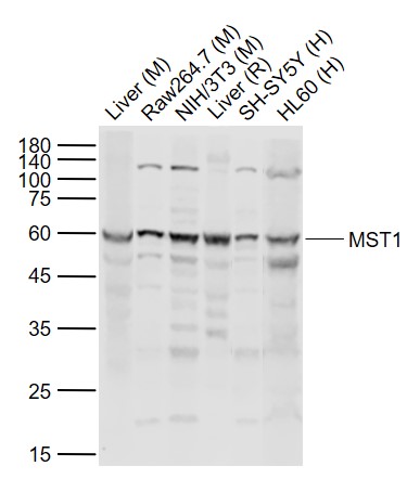
Figure 1. Western blot analysis of MST1/STK4 using anti-MST1/STK4 antibody (A00859-2). Electrophoresis was performed on a 5-20% SDS-PAGE gel at 70V (Stacking gel) / 90V (Resolving gel) for 2-3 hours. The sample well of each lane was loaded with 30 ug of sample under reducing conditions. Lane 1: human Hela whole cell lysates, Lane 2: human HepG2 whole cell lysates, Lane 3: human Jurkat whole cell lysates, Lane 4: human Ramos whole cell lysates, Lane 5: rat liver tissue lysates, Lane 6: mouse spleen tissue lysates. After electrophoresis, proteins were transferred to a nitrocellulose membrane at 150 mA for 50-90 minutes. Blocked the membrane with 5% non-fat milk/TBS for 1.5 hour at RT. The membrane was incubated with rabbit anti-MST1/STK4 antigen affinity purified polyclonal antibody (Catalog # A00859-2) at 0.5 microg/mL overnight at 4°C, then washed with TBS-0.1%Tween 3 times with 5 minutes each and probed with a goat anti-rabbit IgG-HRP secondary antibody at a dilution of 1:5000 for 1.5 hour at RT. The signal is developed using an Enhanced Chemiluminescent detection (ECL) kit (Catalog # EK1002) with Tanon 5200 system. A specific band was detected for MST1/STK4 at approximately 56 kDa. The expected band size for MST1/STK4 is at 56,52 kDa.
Figure 1. Western blot analysis of MST1/STK4 using anti-MST1/STK4 antibody (A00859-2). Electrophoresis was performed on a 5-20% SDS-PAGE gel at 70V (Stacking gel) / 90V (Resolving gel) for 2-3 hours. The sample well of each lane was loaded with 30 ug of sample under reducing conditions. Lane 1: human Hela whole cell lysates, Lane 2: human HepG2 whole cell lysates, Lane 3: human Jurkat whole cell lysates, Lane 4: human Ramos whole cell lysates, Lane 5: rat liver tissue lysates, Lane 6: mouse spleen tissue lysates. After electrophoresis, proteins were transferred to a nitrocellulose membrane at 150 mA for 50-90 minutes. Blocked the membrane with 5% non-fat milk/TBS for 1.5 hour at RT. The membrane was incubated with rabbit anti-MST1/STK4 antigen affinity purified polyclonal antibody (Catalog # A00859-2) at 0.5 microg/mL overnight at 4°C, then washed with TBS-0.1%Tween 3 times with 5 minutes each and probed with a goat anti-rabbit IgG-HRP secondary antibody at a dilution of 1:5000 for 1.5 hour at RT. The signal is developed using an Enhanced Chemiluminescent detection (ECL) kit (Catalog # EK1002) with Tanon 5200 system. A specific band was detected for MST1/STK4 at approximately 56 kDa. The expected band size for MST1/STK4 is at 56,52 kDa.
Anti-MST1/STK4 Antibody Picoband(r)
A00859-2-CARRIER-FREE
ApplicationsWestern Blot, ELISA
Product group Antibodies
ReactivityHuman, Mouse, Rat
TargetSTK4
Overview
- SupplierBoster Bio
- Product NameAnti-MST1/STK4 Antibody Picoband(r)
- Delivery Days Customer9
- ApplicationsWestern Blot, ELISA
- CertificationResearch Use Only
- ClonalityPolyclonal
- Concentration500 ug/ml
- Gene ID6789
- Target nameSTK4
- Target descriptionserine/threonine kinase 4
- Target synonymsKRS2, MST1, YSK3, serine/threonine-protein kinase 4, STE20-like kinase MST1, hippo homolog, kinase responsive to stress 2, mammalian STE20-like protein kinase 1, mammalian sterile 20-like 1, serine/threonine-protein kinase Krs-2
- HostRabbit
- Protein IDQ13043
- Protein NameSerine/threonine-protein kinase 4
- Scientific DescriptionBoster Bio Anti-MST1/STK4 Antibody Picoband® catalog # A00859-2. Tested in WB, ELISA applications. This antibody reacts with Human, Mouse, Rat. The brand Picoband indicates this is a premium antibody that guarantees superior quality, high affinity, and strong signals with minimal background in Western blot applications. Only our best-performing antibodies are designated as Picoband, ensuring unmatched performance.
- ReactivityHuman, Mouse, Rat
- Storage Instruction-20°C,2°C to 8°C
- UNSPSC12352203
Related products
Product group Antibodies
Anti-STK4 Antibody144-08043
ApplicationsImmunoPrecipitation, Western Blot, ImmunoHistoChemistry
ReactivityHuman, Mouse, Rat
TargetSTK4
- SizePrice
Product group Antibodies
MST1 Polyclonal AntibodyBS-28134R
ApplicationsWestern Blot, ELISA
ReactivityBovine, Chicken, Equine, Human, Mouse, Rabbit, Rat
TargetSTK4
- SizePrice
Product group Antibodies
STK4 AntibodyCSB-PA168071
ApplicationsWestern Blot, ELISA, ImmunoHistoChemistry
ReactivityHuman, Mouse
TargetSTK4
- SizePrice
Product group Antibodies

STK4 AntibodyLS-C403027
ApplicationsWestern Blot, ELISA, ImmunoHistoChemistry
ReactivityHuman, Mouse
TargetSTK4
- SizePrice
Product group Antibodies
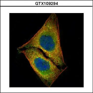
MST1 antibodyGTX109294
ApplicationsImmunoFluorescence, ImmunoPrecipitation, Western Blot, ImmunoCytoChemistry, ImmunoHistoChemistry, ImmunoHistoChemistry Paraffin
ReactivityHuman
TargetSTK4
- SizePrice

Product group Antibodies
Anti-STK4 AntibodyHPA015270
ApplicationsWestern Blot, ImmunoCytoChemistry, ImmunoHistoChemistry
ReactivityHuman, Mouse, Rat
TargetSTK4
- SizePrice
Product group Antibodies
Anti-MST1/STK4 AntibodyCAB8043
ApplicationsImmunoFluorescence, ImmunoPrecipitation, Western Blot, ELISA, ImmunoCytoChemistry, ImmunoHistoChemistry, ImmunoHistoChemistry Paraffin
ReactivityHuman
TargetSTK4
- SizePrice