


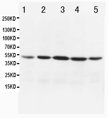
Figure 1. Western blot analysis of MTCO1 using anti-MTCO1 antibody (PA1317-1). Electrophoresis was performed on a 5-20% SDS-PAGE gel at 70V (Stacking gel) / 90V (Resolving gel) for 2-3 hours. The samples were loaded under reducing conditions. Lane 1: human HeLa mitochondria lysates at 20ug, Lane 2: human HeLa whole cell lysates at 20ug, Lane 3: human Caco-2 whole cell lysates at 50ug, Lane 4: rat heart tissue lysates at 50ug. Lane 5: mouse heart tissue lysates at 50ug. After Electrophoresis, proteins were transferred to a Nitrocellulose membrane at 150mA for 60 minutes. Blocked the membrane with 5% Non-fat Milk in TBS for 1.5 hour at RT. The membrane was incubated with rabbit anti-MTCO1 antigen affinity purified polyclonal antibody (Catalog # PA1317-1) at 0.5 microg/mL overnight at 4°C, then washed with TBS-0.1%Tween 3 times with 5 minutes each and probed with a goat anti-rabbit IgG-HRP secondary antibody at a dilution of 1:10000 for 1.5 hour at RT. The signal is developed using an Enhanced Chemiluminescent detection (ECL) kit (Catalog # EK1002) with Tanon 5200 system. A specific band was detected for MTCO1 at approximately 37KD. The expected band size for MTCO1 is at 57KD.
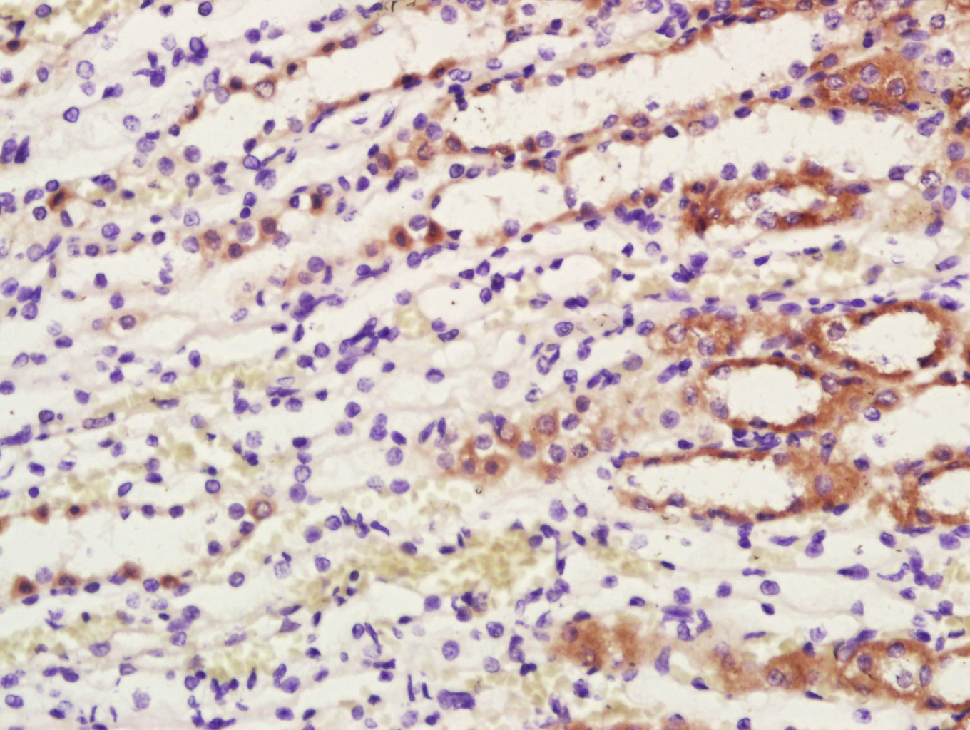
. MTCO1 was detected in paraffin-embedded section of human mammary cancer tissues. Heat mediated antigen retrieval was performed in citrate buffer (pH6, epitope retrieval solution) for 20 mins. The tissue section was blocked with 10% goat serum. The tissue section was then incubated with 1microg/ml rabbit anti-MTCO1 Antibody (PA1317-1) overnight at 4°C. Biotinylated goat anti-rabbit IgG was used as secondary antibody and incubated for 30 minutes at 37°C. The tissue section was developed using Strepavidin-Biotin-Complex (SABC)(Catalog # SA1022) with DAB as the chromogen.")
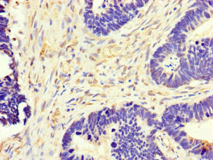
. MTCO1 was detected in immunocytochemical section of C6 Cell. Enzyme antigen retrieval was performed using IHC enzyme antigen retrieval reagent (AR0022) for 15 mins. The cells were blocked with 10% goat serum. And then incubated with 1microg/ml rabbit anti-MTCO1 Antibody (PA1317-1) overnight at 4°C. Biotinylated goat anti-rabbit IgG was used as secondary antibody and incubated for 30 minutes at 37°C. The section was developed using Strepavidin-Biotin-Complex (SABC)(Catalog # SA1022) with DAB as the chromogen.")
Figure 1. Western blot analysis of MTCO1 using anti-MTCO1 antibody (PA1317-1). Electrophoresis was performed on a 5-20% SDS-PAGE gel at 70V (Stacking gel) / 90V (Resolving gel) for 2-3 hours. The samples were loaded under reducing conditions. Lane 1: human HeLa mitochondria lysates at 20ug, Lane 2: human HeLa whole cell lysates at 20ug, Lane 3: human Caco-2 whole cell lysates at 50ug, Lane 4: rat heart tissue lysates at 50ug. Lane 5: mouse heart tissue lysates at 50ug. After Electrophoresis, proteins were transferred to a Nitrocellulose membrane at 150mA for 60 minutes. Blocked the membrane with 5% Non-fat Milk in TBS for 1.5 hour at RT. The membrane was incubated with rabbit anti-MTCO1 antigen affinity purified polyclonal antibody (Catalog # PA1317-1) at 0.5 microg/mL overnight at 4°C, then washed with TBS-0.1%Tween 3 times with 5 minutes each and probed with a goat anti-rabbit IgG-HRP secondary antibody at a dilution of 1:10000 for 1.5 hour at RT. The signal is developed using an Enhanced Chemiluminescent detection (ECL) kit (Catalog # EK1002) with Tanon 5200 system. A specific band was detected for MTCO1 at approximately 37KD. The expected band size for MTCO1 is at 57KD.
Anti-MTCO1/MT-CO1 Antibody Picoband(r)
PA1317-1-CARRIER-FREE
ApplicationsWestern Blot, ImmunoCytoChemistry, ImmunoHistoChemistry
Product group Antibodies
ReactivityHuman
TargetCOX1
Overview
- SupplierBoster Bio
- Product NameAnti-MTCO1/MT-CO1 Antibody Picoband(r)
- Delivery Days Customer9
- Application Supplier NoteTested Species: In-house tested species with positive results. Predicted Species: Species predicted to be fit for the product based on sequence similarities. Other applications have not been tested. Optimal dilutions should be determined by end users.
- ApplicationsWestern Blot, ImmunoCytoChemistry, ImmunoHistoChemistry
- CertificationResearch Use Only
- ClonalityPolyclonal
- Concentration500 ug/ml
- Gene ID4512
- Target nameCOX1
- Target descriptioncytochrome c oxidase subunit I
- Target synonymsCOI, MTCO1, cytochrome c oxidase subunit I
- HostRabbit
- IsotypeIgG
- Protein IDP00395
- Protein NameCytochrome c oxidase subunit 1
- Scientific DescriptionBoster Bio Anti-MTCO1/MT-CO1 Antibody catalog # PA1317-1. Tested in IHC, ICC, WB applications. This antibody reacts with Human. The brand Picoband indicates this is a premium antibody that guarantees superior quality, high affinity, and strong signals with minimal background in Western blot applications. Only our best-performing antibodies are designated as Picoband, ensuring unmatched performance.
- ReactivityHuman
- Storage Instruction-20°C,2°C to 8°C
- UNSPSC12352203
Related products
Product group Antibodies
ApplicationsWestern Blot, ImmunoCytoChemistry
ReactivityHuman, Mouse
- SizePrice
Product group Antibodies
Anti-MTCO1 Antibody144-65228
ApplicationsImmunoFluorescence, Western Blot, ImmunoHistoChemistry
ReactivityHuman, Mouse, Rat
TargetCOX1
- SizePrice
Product group Antibodies

MT-CO1 / COX1 AntibodyLS-C668936
ApplicationsWestern Blot
ReactivityHuman
TargetCOX1
- SizePrice
Product group Antibodies
References
COX1/MTCO1 Polyclonal AntibodyBS-3953R
ApplicationsImmunoFluorescence, Western Blot, ELISA, ImmunoCytoChemistry, ImmunoHistoChemistry, ImmunoHistoChemistry Frozen, ImmunoHistoChemistry Paraffin
ReactivityBovine, Canine, Chicken, Equine, Human, Mouse, Porcine, Rabbit, Rat
TargetCOX1
- SizePrice
Product group Antibodies
MT-CO1 AntibodyCSB-PA015072LA01HU
ApplicationsImmunoFluorescence, ELISA, ImmunoHistoChemistry
ReactivityHuman
TargetCOX1
- SizePrice

Product group Antibodies
Anti-MT-CO1 AntibodyHPA069328
ApplicationsWestern Blot, ImmunoHistoChemistry
ReactivityHuman
TargetCOX1
- SizePrice
Product group Antibodies
MTCO1 antibodyGTX11619
ApplicationsWestern Blot
ReactivityHuman
TargetCOX1
- SizePrice
Product group Antibodies
Anti-MTCO1 AntibodyCAB17889
ApplicationsImmunoFluorescence, Western Blot, ELISA, ImmunoCytoChemistry, ImmunoHistoChemistry, ImmunoHistoChemistry Paraffin
ReactivityHuman
TargetCOX1
- SizePrice