


Figure 1. Western blot analysis of Myosin using anti-Myosin antibody (MA1064). Electrophoresis was performed on a 5-20% SDS-PAGE gel at 70V (Stacking gel) / 90V (Resolving gel) for 2-3 hours. The sample well of each lane was loaded with 30 ug of sample under reducing conditions. Lane 1: monkey heart tissue lysates, Lane 2: monkey skeletal muscle tissue lysates, Lane 3: rat heart tissue lysates, Lane 4: rat skeletal muscle tissue lysates, Lane 5: mouse heart tissue lysates, Lane 6: mouse skeletal muscle tissue lysates. After electrophoresis, proteins were transferred to a nitrocellulose membrane at 150 mA for 50-90 minutes. Blocked the membrane with 5% non-fat milk/TBS for 1.5 hour at RT. The membrane was incubated with mouse anti-Myosin antigen affinity purified monoclonal antibody (Catalog # MA1064) at 0.5 microg/mL overnight at 4°C, then washed with TBS-0.1%Tween 3 times with 5 minutes each and probed with a goat anti-mouse IgG-HRP secondary antibody at a dilution of 1:10000 for 1.5 hour at RT. The signal is developed using an Enhanced Chemiluminescent detection (ECL) kit (Catalog # EK1001) with Tanon 5200 system. A specific band was detected for Myosin at approximately 230 kDa. The expected band size for Myosin is at 223 kDa.
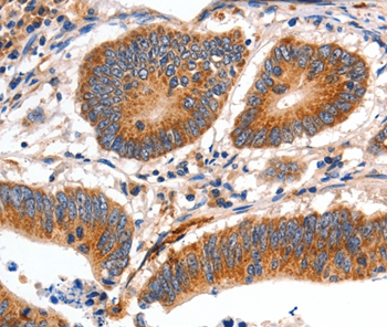
. Myosin was detected in a paraffin-embedded section of rat skeletal muscle tissue. Heat mediated antigen retrieval was performed in EDTA buffer (pH 8.0, epitope retrieval solution). The tissue section was blocked with 10% goat serum. The tissue section was then incubated with 2 microg/ml mouse anti-Myosin Antibody (MA1064) overnight at 4°C. Peroxidase Conjugated Goat Anti-mouse IgG was used as secondary antibody and incubated for 30 minutes at 37°C. The tissue section was developed using HRP Conjugated Mouse IgG Super Vision Assay Kit (Catalog # SV0001) with DAB as the chromogen.")
Figure 1. Western blot analysis of Myosin using anti-Myosin antibody (MA1064). Electrophoresis was performed on a 5-20% SDS-PAGE gel at 70V (Stacking gel) / 90V (Resolving gel) for 2-3 hours. The sample well of each lane was loaded with 30 ug of sample under reducing conditions. Lane 1: monkey heart tissue lysates, Lane 2: monkey skeletal muscle tissue lysates, Lane 3: rat heart tissue lysates, Lane 4: rat skeletal muscle tissue lysates, Lane 5: mouse heart tissue lysates, Lane 6: mouse skeletal muscle tissue lysates. After electrophoresis, proteins were transferred to a nitrocellulose membrane at 150 mA for 50-90 minutes. Blocked the membrane with 5% non-fat milk/TBS for 1.5 hour at RT. The membrane was incubated with mouse anti-Myosin antigen affinity purified monoclonal antibody (Catalog # MA1064) at 0.5 microg/mL overnight at 4°C, then washed with TBS-0.1%Tween 3 times with 5 minutes each and probed with a goat anti-mouse IgG-HRP secondary antibody at a dilution of 1:10000 for 1.5 hour at RT. The signal is developed using an Enhanced Chemiluminescent detection (ECL) kit (Catalog # EK1001) with Tanon 5200 system. A specific band was detected for Myosin at approximately 230 kDa. The expected band size for Myosin is at 223 kDa.
Anti-Myosin(Skeletal, Slow) Myh7 Antibody (Monoclonal, NOQ7.5.4D)
MA1064
ApplicationsWestern Blot, ImmunoHistoChemistry
Product group Antibodies
ReactivityHuman, Monkey, Mouse, Rat
TargetMYH7
Overview
- SupplierBoster Bio
- Product NameAnti-Myosin(Skeletal, Slow) Myh7 Antibody (Monoclonal, NOQ7.5.4D)
- Delivery Days Customer9
- Application Supplier NoteOther applications have not been tested. Optimal dilutions should be determined by end users.
- ApplicationsWestern Blot, ImmunoHistoChemistry
- Applications SupplierIHP, WB, IHC
- CertificationResearch Use Only
- ClonalityMonoclonal
- Clone IDNOQ7.5.4D
- Concentration100 ug/ml
- Gene ID4625
- Target nameMYH7
- Target descriptionmyosin heavy chain 7
- Target synonymsCMD1S, CMH1, CMYO7A, CMYO7B, CMYP7A, CMYP7B, MPD1, MYHCB, SPMD, SPMM, myosin-7, cardiac muscle myosin heavy chain 7 beta, myHC-beta, myhc-slow, myosin 7, myosin heavy chain beta-subunit, myosin, heavy chain 7, cardiac muscle, beta, myosin, heavy polypeptide 7, cardiac muscle, beta, rhabdomyosarcoma antigen MU-RMS-40.7A
- HostMouse
- IsotypeIgG1
- Protein IDP12883
- Protein NameMyosin-7
- Scientific DescriptionBoster Bio Anti-Myosin (Skeletal, Slow) Myh7 Antibody (Monoclonal, NOQ7.5.4D) catalog # MA1064. Tested in IHC, WB applications. This antibody reacts with Human, Monkey, Mouse, Rat.
- ReactivityHuman, Monkey, Mouse, Rat
- Reactivity SupplierHuman, Mouse, Rabbit, Rat
- Storage Instruction-20°C,2°C to 8°C
- UNSPSC12352203
References
- Ma X, Chang H, Wang Z, et al. Differential activation of the calpain system involved in individualized adaptation of different fast-twitch muscles in hibernating Daurian ground squirrels. J Appl Physiol (1985). 2019,127(2):328-341. doi: 10.1152/japplphysiol.00124.2019Read this paper
- Wang LP, Fan SJ, Li SM, et al. Oxidative stress promotes myocardial fibrosis by upregulating K(Ca)3.1 channel expression in AGT-REN double transgenic hypertensive mice. Pflugers Arch. 2017,469(9):1061-1071. doi: 10.1007/s00424-017-1984-0Read this paper
Datasheet
MSDS
Related products
Product group Antibodies
Anti-MYH7 AntibodyA46718
ApplicationsImmunoHistoChemistry
ReactivityHuman
- SizePrice
Product group Antibodies
Anti-MYH7 Antibody144-07564
ApplicationsWestern Blot, ImmunoHistoChemistry
ReactivityHuman, Mouse, Rat
TargetMYH7
- SizePrice
Product group Antibodies

MYH7 AntibodyLS-C668921
ApplicationsWestern Blot, ImmunoHistoChemistry, ImmunoHistoChemistry Paraffin
ReactivityHuman
TargetMYH7
- SizePrice
Product group Antibodies
References
ApplicationsFlow Cytometry, ImmunoFluorescence, ELISA, ImmunoCytoChemistry, ImmunoHistoChemistry, ImmunoHistoChemistry Frozen, ImmunoHistoChemistry Paraffin
ReactivityBovine, Canine, Equine, Human, Mouse, Porcine, Rabbit, Rat, Sheep
TargetMYH7
- SizePrice
Product group Antibodies
ApplicationsImmunoPrecipitation, Western Blot, ImmunoCytoChemistry, ImmunoHistoChemistry
ReactivityPorcine
TargetMYH7
- SizePrice
Product group Antibodies
MYH7 AntibodyCSB-PA015300LA01HU
ApplicationsELISA, ImmunoHistoChemistry
ReactivityHuman
TargetMYH7
- SizePrice
![Mouse tissue extract (50 μg) was separated by 5% SDS-PAGE, and the membrane was blotted with MYH7 antibody [N1], N-term (GTX100713) diluted at 1:2000.](https://www.genetex.com/upload/website/prouct_img/normal/GTX100713/GTX100713_40058_20160505_WB_M_muscle_w_23060100_417.webp)
Product group Antibodies
MYH7 antibody [N1], N-termGTX100713
ApplicationsWestern Blot
ReactivityHuman, Mouse, Rat
TargetMYH7
- SizePrice

Product group Antibodies
Anti-MYH7 AntibodyHPA001239
ApplicationsImmunoHistoChemistry
ReactivityHuman
TargetMYH7
- SizePrice
Product group Antibodies
Anti-MYH7/beta-MHC AntibodyCAB7564
ApplicationsImmunoFluorescence, Western Blot, ELISA, ImmunoCytoChemistry
ReactivityMouse
TargetMYH7
- SizePrice