

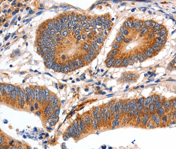
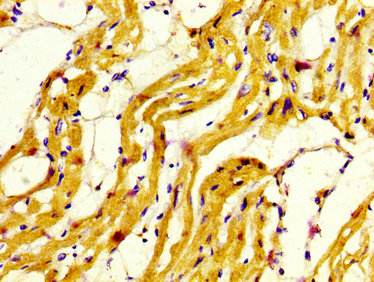
Myosin Heavy Chain 7, Cardiac Muscle, Beta (MYH7) Polyclonal Antibody
CAU23356
ApplicationsImmunoPrecipitation, Western Blot, ImmunoCytoChemistry, ImmunoHistoChemistry
Product group Antibodies
ReactivityPorcine
TargetMYH7
Overview
- SupplierBiomatik
- Product NameMyosin Heavy Chain 7, Cardiac Muscle, Beta (MYH7) Polyclonal Antibody
- Delivery Days Customer12
- ApplicationsImmunoPrecipitation, Western Blot, ImmunoCytoChemistry, ImmunoHistoChemistry
- Applications SupplierWB; IHC; ICC; IP.
- CertificationResearch Use Only
- ClonalityPolyclonal
- Concentration0.5 mg/ml
- ConjugateUnconjugated
- Gene ID4625
- Target nameMYH7
- Target descriptionmyosin heavy chain 7
- Target synonymsCMD1S, CMH1, CMYO7A, CMYO7B, CMYP7A, CMYP7B, MPD1, MYHCB, SPMD, SPMM, myosin-7, cardiac muscle myosin heavy chain 7 beta, myHC-beta, myhc-slow, myosin 7, myosin heavy chain beta-subunit, myosin, heavy chain 7, cardiac muscle, beta, myosin, heavy polypeptide 7, cardiac muscle, beta, rhabdomyosarcoma antigen MU-RMS-40.7A
- HostRabbit
- Protein IDP12883
- Protein NameMyosin-7
- Scientific DescriptionThe Myosin Heavy Chain 7, Cardiac Muscle, Beta (MYH7) Polyclonal Antibody (Species: Human) has been validated for the following applications: WB, IHC, ICC, IP.
- ReactivityPorcine
- Reactivity SupplierHuman
- Storage Instruction-20°C,2°C to 8°C
- UNSPSC12352203
Related products
Product group Antibodies
Anti-MYH7 AntibodyA46718
ApplicationsImmunoHistoChemistry
ReactivityHuman
- SizePrice
Product group Antibodies
Anti-MYH7 Antibody144-07564
ApplicationsWestern Blot, ImmunoHistoChemistry
ReactivityHuman, Mouse, Rat
TargetMYH7
- SizePrice

Product group Antibodies
References
ApplicationsWestern Blot, ImmunoHistoChemistry
ReactivityHuman, Monkey, Mouse, Rat
TargetMYH7
- SizePrice
Product group Antibodies

MYH7 AntibodyLS-C668921
ApplicationsWestern Blot, ImmunoHistoChemistry, ImmunoHistoChemistry Paraffin
ReactivityHuman
TargetMYH7
- SizePrice
Product group Antibodies
References
ApplicationsFlow Cytometry, ImmunoFluorescence, ELISA, ImmunoCytoChemistry, ImmunoHistoChemistry, ImmunoHistoChemistry Frozen, ImmunoHistoChemistry Paraffin
ReactivityBovine, Canine, Equine, Human, Mouse, Porcine, Rabbit, Rat, Sheep
TargetMYH7
- SizePrice
Product group Antibodies
MYH7 AntibodyCSB-PA015300LA01HU
ApplicationsELISA, ImmunoHistoChemistry
ReactivityHuman
TargetMYH7
- SizePrice
![Mouse tissue extract (50 μg) was separated by 5% SDS-PAGE, and the membrane was blotted with MYH7 antibody [N1], N-term (GTX100713) diluted at 1:2000.](https://www.genetex.com/upload/website/prouct_img/normal/GTX100713/GTX100713_40058_20160505_WB_M_muscle_w_23060100_417.webp)
Product group Antibodies
MYH7 antibody [N1], N-termGTX100713
ApplicationsWestern Blot
ReactivityHuman, Mouse, Rat
TargetMYH7
- SizePrice

Product group Antibodies
Anti-MYH7 AntibodyHPA001239
ApplicationsImmunoHistoChemistry
ReactivityHuman
TargetMYH7
- SizePrice
Product group Antibodies
Anti-MYH7/beta-MHC AntibodyCAB7564
ApplicationsImmunoFluorescence, Western Blot, ELISA, ImmunoCytoChemistry
ReactivityMouse
TargetMYH7
- SizePrice