

MYH7 Antibody
LS-C668921
ApplicationsWestern Blot, ImmunoHistoChemistry, ImmunoHistoChemistry Paraffin
Product group Antibodies
ReactivityHuman
TargetMYH7
Overview
- SupplierLifeSpan BioSciences
- Product NameMYH7 Antibody
- Delivery Days Customer23
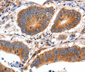
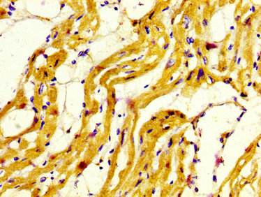
- Application Supplier NoteWestern blot analysis of Myosin 7 expression in mouse skeletal muscle (A), rat skeletal muscle (B) whole cell lysates. Immunohistochemical analysis of Myosin 7 staining in human lung cancer formalin fixed paraffin embedded tissue section. The section was pre-treated using heat mediated antigen retrieval with sodium citrate buffer (pH 6.0). The section was then incubated with the antibody at room temperature and detected using an HRP conjugated compact polymer system. DAB was used as the chromogen. The section was then counterstained with haematoxylin and mounted with DPX.. IHC (1:50 - 1:200), IHC-P, WB (1:500 - 1:2000) Western blot analysis of Myosin 7 expression in mouse skeletal muscle (A), rat skeletal muscle (B) whole cell lysates. Immunohistochemical analysis of Myosin 7 staining in human lung cancer formalin fixed paraffin embedded tissue section. The section was pre-treated using heat mediated antigen retrieval with sodium citrate buffer (pH 6.0). The section was then incubated with the antibody at room temperature and detected using an HRP conjugated compact polymer system. DAB was used as the chromogen. The section was then counterstained with haematoxylin and mounted with DPX.
- ApplicationsWestern Blot, ImmunoHistoChemistry, ImmunoHistoChemistry Paraffin
- CertificationResearch Use Only
- ClonalityPolyclonal
- Concentration1 mg/ml
- ConjugateUnconjugated
- Gene ID4625
- Target nameMYH7
- Target descriptionmyosin heavy chain 7
- Target synonymsCMD1S, CMH1, CMYO7A, CMYO7B, CMYP7A, CMYP7B, MPD1, MYHCB, SPMD, SPMM, myosin-7, cardiac muscle myosin heavy chain 7 beta, myHC-beta, myhc-slow, myosin 7, myosin heavy chain beta-subunit, myosin, heavy chain 7, cardiac muscle, beta, myosin, heavy polypeptide 7, cardiac muscle, beta, rhabdomyosarcoma antigen MU-RMS-40.7A
- HostRabbit
- ReactivityHuman
- Storage Instruction-20°C
- UNSPSC41116161
Related products
Product group Antibodies
Anti-MYH7 AntibodyA46718
ApplicationsImmunoHistoChemistry
ReactivityHuman
- SizePrice
Product group Antibodies
Anti-MYH7 Antibody144-07564
ApplicationsWestern Blot, ImmunoHistoChemistry
ReactivityHuman, Mouse, Rat
TargetMYH7
- SizePrice

Product group Antibodies
References
ApplicationsWestern Blot, ImmunoHistoChemistry
ReactivityHuman, Monkey, Mouse, Rat
TargetMYH7
- SizePrice
Product group Antibodies
References
ApplicationsFlow Cytometry, ImmunoFluorescence, ELISA, ImmunoCytoChemistry, ImmunoHistoChemistry, ImmunoHistoChemistry Frozen, ImmunoHistoChemistry Paraffin
ReactivityBovine, Canine, Equine, Human, Mouse, Porcine, Rabbit, Rat, Sheep
TargetMYH7
- SizePrice
Product group Antibodies
ApplicationsImmunoPrecipitation, Western Blot, ImmunoCytoChemistry, ImmunoHistoChemistry
ReactivityPorcine
TargetMYH7
- SizePrice
Product group Antibodies
MYH7 AntibodyCSB-PA015300LA01HU
ApplicationsELISA, ImmunoHistoChemistry
ReactivityHuman
TargetMYH7
- SizePrice
![Mouse tissue extract (50 μg) was separated by 5% SDS-PAGE, and the membrane was blotted with MYH7 antibody [N1], N-term (GTX100713) diluted at 1:2000.](https://www.genetex.com/upload/website/prouct_img/normal/GTX100713/GTX100713_40058_20160505_WB_M_muscle_w_23060100_417.webp)
Product group Antibodies
MYH7 antibody [N1], N-termGTX100713
ApplicationsWestern Blot
ReactivityHuman, Mouse, Rat
TargetMYH7
- SizePrice

Product group Antibodies
Anti-MYH7 AntibodyHPA001239
ApplicationsImmunoHistoChemistry
ReactivityHuman
TargetMYH7
- SizePrice
Product group Antibodies
Anti-MYH7/beta-MHC AntibodyCAB7564
ApplicationsImmunoFluorescence, Western Blot, ELISA, ImmunoCytoChemistry
ReactivityMouse
TargetMYH7
- SizePrice