

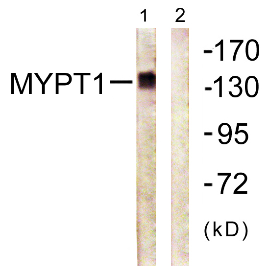


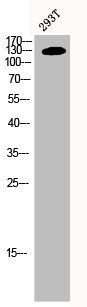
Anti-MYPT1 Antibody
A95830
ApplicationsWestern Blot, ELISA, ImmunoHistoChemistry
Product group Antibodies
ReactivityHuman, Mouse, Rat
Overview
- SupplierAntibodies.com
- Product NameAnti-MYPT1 Antibody
- Delivery Days Customer7
- ApplicationsWestern Blot, ELISA, ImmunoHistoChemistry
- CertificationResearch Use Only
- ClonalityPolyclonal
- ConjugateUnconjugated
- HostRabbit
- IsotypeIgG
- Scientific DescriptionRabbit polyclonal antibody to MYPT1.
- ReactivityHuman, Mouse, Rat
- UNSPSC12352203
Related products
Product group Antibodies
Anti-PPP1R12A Antibody144-00587
ApplicationsImmunoPrecipitation, Western Blot
ReactivityHuman, Mouse
TargetPPP1R12A
- SizePrice
Product group Antibodies
ApplicationsFlow Cytometry, Western Blot
ReactivityBovine, Canine, Chicken, Equine, Human, Mouse, Rabbit, Rat
TargetPPP1R12A
- SizePrice
Product group Antibodies
PPP1R12A AntibodyCSB-PA003352
ApplicationsWestern Blot, ELISA, ImmunoHistoChemistry
ReactivityHuman, Monkey, Mouse, Rat
TargetPPP1R12A
- SizePrice
Product group Antibodies

PPP1R12A / MYPT1 AntibodyLS-C401866
ApplicationsWestern Blot, ELISA
ReactivityHuman, Mouse, Rat
TargetPPP1R12A
- SizePrice

Product group Antibodies
Anti-PPP1R12A AntibodyHPA041296
ApplicationsImmunoCytoChemistry
ReactivityHuman
TargetPPP1R12A
- SizePrice
Product group Antibodies
MYPT1 antibodyGTX112344
ApplicationsImmunoFluorescence, ImmunoCytoChemistry
ReactivityHuman
TargetPPP1R12A
- SizePrice
Product group Antibodies
Anti-PPP1R12A AntibodyCAB0587
ApplicationsImmunoFluorescence, ImmunoPrecipitation, Western Blot, ELISA, ImmunoCytoChemistry
ReactivityHuman
TargetPPP1R12A
- SizePrice

Product group Antibodies
Anti-Myosin Phosphatase/PPP1R12A Antibody Picoband(r)PB9737-CARRIER-FREE
ApplicationsFlow Cytometry, ImmunoFluorescence, Western Blot, ImmunoCytoChemistry, ImmunoHistoChemistry
ReactivityBovine, Canine, Chicken, Equine, Human, Monkey, Mouse, Rabbit, Rat
TargetPPP1R12A
- SizePrice