

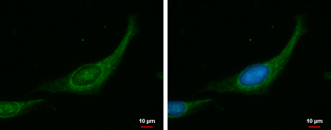
MYPT1 antibody detects MYPT1 protein at cytoplasm by immunofluorescent analysis. Sample: RD cells were fixed in ice-cold MeOH for 5 min. Green: MYPT1 protein stained by MYPT1 antibody (GTX112344) diluted at 1:1000. Blue: Hoechst 33342 staining.
MYPT1 antibody detects MYPT1 protein at cytoplasm by immunofluorescent analysis. Sample: RD cells were fixed in ice-cold MeOH for 5 min. Green: MYPT1 protein stained by MYPT1 antibody (GTX112344) diluted at 1:1000. Blue: Hoechst 33342 staining.
MYPT1 antibody
GTX112344
ApplicationsImmunoFluorescence, ImmunoCytoChemistry
Product group Antibodies
ReactivityHuman
TargetPPP1R12A
Overview
- SupplierGeneTex
- Product NameMYPT1 antibody
- Delivery Days Customer9
- Application Supplier NoteICC/IF: 1:100-1:1000. *Optimal dilutions/concentrations should be determined by the researcher.Not tested in other applications.
- ApplicationsImmunoFluorescence, ImmunoCytoChemistry
- CertificationResearch Use Only
- ClonalityPolyclonal
- Concentration1 mg/ml
- ConjugateUnconjugated
- Gene ID4659
- Target namePPP1R12A
- Target descriptionprotein phosphatase 1 regulatory subunit 12A
- Target synonymsGUBS, M130, MBS, MYPT1, protein phosphatase 1 regulatory subunit 12A, myosin binding subunit, myosin phosphatase, target subunit 1, myosin phosphatase-targeting subunit 1, protein phosphatase 1, regulatory (inhibitor) subunit 12A, protein phosphatase myosin-binding subunit
- HostRabbit
- IsotypeIgG
- Protein IDO14974
- Protein NameProtein phosphatase 1 regulatory subunit 12A
- Scientific DescriptionMyosin phosphatase target subunit 1, which is also called the myosin-binding subunit of myosin phosphatase, is one of the subunits of myosin phosphatase. Myosin phosphatase regulates the interaction of actin and myosin downstream of the guanosine triphosphatase Rho. The small guanosine triphosphatase Rho is implicated in myosin light chain (MLC) phosphorylation, which results in contraction of smooth muscle and interaction of actin and myosin in nonmuscle cells. The guanosine triphosphate (GTP)-bound, active form of RhoA (GTP.RhoA) specifically interacted with the myosin-binding subunit (MBS) of myosin phosphatase, which regulates the extent of phosphorylation of MLC. Rho-associated kinase (Rho-kinase), which is activated by GTP. RhoA, phosphorylated MBS and consequently inactivated myosin phosphatase. Overexpression of RhoA or activated RhoA in NIH 3T3 cells increased phosphorylation of MBS and MLC. Thus, Rho appears to inhibit myosin phosphatase through the action of Rho-kinase. Several transcript variants encoding different isoforms have been found for this gene. [provided by RefSeq]
- ReactivityHuman
- Storage Instruction-20°C or -80°C,2°C to 8°C
- UNSPSC41116161
Datasheet
Related products
Product group Antibodies
Anti-MYPT1 AntibodyA95830
ApplicationsWestern Blot, ELISA, ImmunoHistoChemistry
ReactivityHuman, Mouse, Rat
- SizePrice
Product group Antibodies
Anti-PPP1R12A Antibody144-00587
ApplicationsImmunoPrecipitation, Western Blot
ReactivityHuman, Mouse
TargetPPP1R12A
- SizePrice
Product group Antibodies
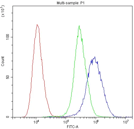
ApplicationsFlow Cytometry, Western Blot
ReactivityBovine, Canine, Chicken, Equine, Human, Mouse, Rabbit, Rat
TargetPPP1R12A
- SizePrice
Product group Antibodies
PPP1R12A AntibodyCSB-PA003352
ApplicationsWestern Blot, ELISA, ImmunoHistoChemistry
ReactivityHuman, Monkey, Mouse, Rat
TargetPPP1R12A
- SizePrice
Product group Antibodies

PPP1R12A / MYPT1 AntibodyLS-C401866
ApplicationsWestern Blot, ELISA
ReactivityHuman, Mouse, Rat
TargetPPP1R12A
- SizePrice
Product group Antibodies
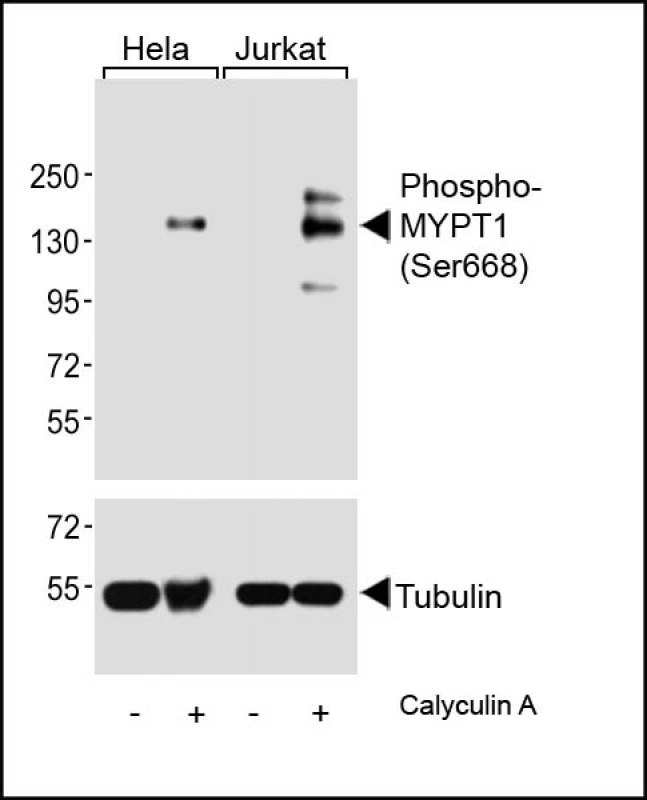
MYPT1 (phospho Ser668) antibodyGTX00776
ApplicationsWestern Blot
ReactivityHuman
TargetPPP1R12A
- SizePrice

Product group Antibodies
Anti-PPP1R12A AntibodyHPA041296
ApplicationsImmunoCytoChemistry
ReactivityHuman
TargetPPP1R12A
- SizePrice
Product group Antibodies
MYPT1 antibodyGTX12027
ApplicationsFlow Cytometry, Western Blot
ReactivityHuman, Mouse, Rat
TargetPPP1R12A
- SizePrice
Product group Antibodies
Anti-PPP1R12A AntibodyCAB0587
ApplicationsImmunoFluorescence, ImmunoPrecipitation, Western Blot, ELISA, ImmunoCytoChemistry
ReactivityHuman
TargetPPP1R12A
- SizePrice