


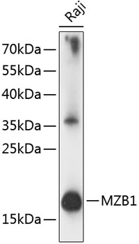
Figure 1. Western blot analysis of MZB1 using anti-MZB1 antibody (A08281-1). Electrophoresis was performed on a 5-20% SDS-PAGE gel at 70V (Stacking gel) / 90V (Resolving gel) for 2-3 hours. The sample well of each lane was loaded with 30 ug of sample under reducing conditions. Lane 1: human Raji whole cell lysates, Lane 2: human Jurkat whole cell lysates. After electrophoresis, proteins were transferred to a nitrocellulose membrane at 150 mA for 50-90 minutes. Blocked the membrane with 5% non-fat milk/TBS for 1.5 hour at RT. The membrane was incubated with rabbit anti-MZB1 antigen affinity purified polyclonal antibody (Catalog # A08281-1) at 0.25 microg/mL overnight at 4°C, then washed with TBS-0.1%Tween 3 times with 5 minutes each and probed with a goat anti-rabbit IgG-HRP secondary antibody at a dilution of 1:5000 for 1.5 hour at RT. The signal is developed using an Enhanced Chemiluminescent detection (ECL) kit (Catalog # EK1002) with Tanon 5200 system. A specific band was detected for MZB1 at approximately 19 kDa. The expected band size for MZB1 is at 21 kDa.
. MZB1 was detected in a paraffin-embedded section of human spleen tissue. Heat mediated antigen retrieval was performed in EDTA buffer (pH 8.0, epitope retrieval solution). The tissue section was blocked with 10% goat serum. The tissue section was then incubated with 2 microg/ml rabbit anti-MZB1 Antibody (A08281-1) overnight at 4°C. Peroxidase Conjugated Goat Anti-rabbit IgG was used as secondary antibody and incubated for 30 minutes at 37°C. The tissue section was developed using HRP Conjugated Rabbit IgG Super Vision Assay Kit (Catalog # SV0002) with DAB as the chromogen.")
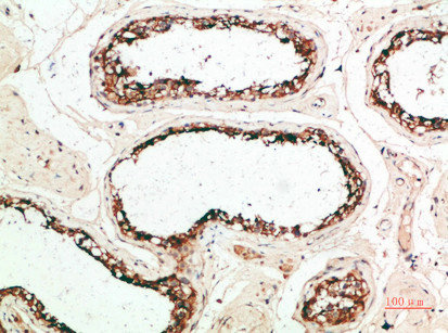
. MZB1 was detected in a paraffin-embedded section of human tonsil tissue. Heat mediated antigen retrieval was performed in EDTA buffer (pH 8.0, epitope retrieval solution). The tissue section was blocked with 10% goat serum. The tissue section was then incubated with 2 microg/ml rabbit anti-MZB1 Antibody (A08281-1) overnight at 4°C. Peroxidase Conjugated Goat Anti-rabbit IgG was used as secondary antibody and incubated for 30 minutes at 37°C. The tissue section was developed using HRP Conjugated Rabbit IgG Super Vision Assay Kit (Catalog # SV0002) with DAB as the chromogen.")
. MZB1 was detected in a paraffin-embedded section of human tonsil tissue. Heat mediated antigen retrieval was performed in EDTA buffer (pH 8.0, epitope retrieval solution). The tissue section was blocked with 10% goat serum. The tissue section was then incubated with 2 microg/ml rabbit anti-MZB1 Antibody (A08281-1) overnight at 4°C. Peroxidase Conjugated Goat Anti-rabbit IgG was used as secondary antibody and incubated for 30 minutes at 37°C. The tissue section was developed using HRP Conjugated Rabbit IgG Super Vision Assay Kit (Catalog # SV0002) with DAB as the chromogen.")
. MZB1 was detected in an immunocytochemical section of Jurkat cells. Enzyme antigen retrieval was performed using IHC enzyme antigen retrieval reagent (AR0022) for 15 mins. The cells were blocked with 10% goat serum. And then incubated with 5 microg/mL rabbit anti-MZB1 Antibody (A08281-1) overnight at 4°C. DyLight®488 Conjugated Goat Anti-Rabbit IgG (BA1127) was used as secondary antibody at 1:500 dilution and incubated for 30 minutes at 37°C. The section was counterstained with DAPI. Visualize using a fluorescence microscope and filter sets appropriate for the label used.")
. Overlay histogram showing JK cells stained with A08281-1 (Blue line). To facilitate intracellular staining, cells were fixed with 4% paraformaldehyde and permeabilized with permeabilization buffer. The cells were blocked with 10% normal goat serum. And then incubated with rabbit anti-MZB1 Antibody (A08281-1, 1 microg/1x106 cells) for 30 min at 20°C. DyLight®488 conjugated goat anti-rabbit IgG (BA1127, 5-10 microg/1x106 cells) was used as secondary antibody for 30 minutes at 20°C. Isotype control antibody (Green line) was rabbit IgG (1 microg/1x106) used under the same conditions. Unlabelled sample (Red line) was also used as a control.")
Figure 1. Western blot analysis of MZB1 using anti-MZB1 antibody (A08281-1). Electrophoresis was performed on a 5-20% SDS-PAGE gel at 70V (Stacking gel) / 90V (Resolving gel) for 2-3 hours. The sample well of each lane was loaded with 30 ug of sample under reducing conditions. Lane 1: human Raji whole cell lysates, Lane 2: human Jurkat whole cell lysates. After electrophoresis, proteins were transferred to a nitrocellulose membrane at 150 mA for 50-90 minutes. Blocked the membrane with 5% non-fat milk/TBS for 1.5 hour at RT. The membrane was incubated with rabbit anti-MZB1 antigen affinity purified polyclonal antibody (Catalog # A08281-1) at 0.25 microg/mL overnight at 4°C, then washed with TBS-0.1%Tween 3 times with 5 minutes each and probed with a goat anti-rabbit IgG-HRP secondary antibody at a dilution of 1:5000 for 1.5 hour at RT. The signal is developed using an Enhanced Chemiluminescent detection (ECL) kit (Catalog # EK1002) with Tanon 5200 system. A specific band was detected for MZB1 at approximately 19 kDa. The expected band size for MZB1 is at 21 kDa.
Anti-MZB1 Antibody Picoband(r)
A08281-1-CARRIER-FREE
ApplicationsFlow Cytometry, ImmunoFluorescence, Western Blot, ELISA, ImmunoCytoChemistry, ImmunoHistoChemistry
Product group Antibodies
ReactivityHuman
TargetMZB1
Overview
- SupplierBoster Bio
- Product NameAnti-MZB1 Antibody Picoband(r)
- Delivery Days Customer9
- ApplicationsFlow Cytometry, ImmunoFluorescence, Western Blot, ELISA, ImmunoCytoChemistry, ImmunoHistoChemistry
- CertificationResearch Use Only
- ClonalityPolyclonal
- Concentration500 ug/ml
- Gene ID51237
- Target nameMZB1
- Target descriptionmarginal zone B and B1 cell specific protein
- Target synonymsMEDA-7, PACAP, pERp1, marginal zone B- and B1-cell-specific protein, HSPC190, caspase-2 binding protein, mesenteric estrogen-dependent adipose 7, mesenteric oestrogen-dependent adipose gene- 7, plasma cell-induced ER protein 1, plasma cell-induced resident ER protein, plasma cell-induced resident endoplasmic reticulum protein, proapoptotic caspase adapter protein, proapoptotic caspase adaptor protein
- HostRabbit
- IsotypeIgG
- Protein IDQ8WU39
- Protein NameMarginal zone B- and B1-cell-specific protein
- Scientific DescriptionBoster Bio Anti-MZB1 Antibody Picoband® catalog # A08281-1. Tested in ELISA, IF, IHC, ICC, WB, Flow Cytometry applications. This antibody reacts with Human. The brand Picoband indicates this is a premium antibody that guarantees superior quality, high affinity, and strong signals with minimal background in Western blot applications. Only our best-performing antibodies are designated as Picoband, ensuring unmatched performance.
- ReactivityHuman
- Storage Instruction-20°C,2°C to 8°C
- UNSPSC12352203
Related products
Product group Antibodies
Anti-PACAP AntibodyA88543
ApplicationsImmunoFluorescence, Western Blot, ImmunoCytoChemistry, ImmunoHistoChemistry
ReactivityHuman, Mouse, Rat
- SizePrice
Product group Antibodies
Anti-MZB1 Antibody144-63304
ApplicationsImmunoFluorescence, Western Blot, ImmunoHistoChemistry
ReactivityHuman, Mouse, Rat
TargetMZB1
- SizePrice
Product group Antibodies

ApplicationsELISA
ReactivityHuman
TargetMZB1
- SizePrice
Product group Antibodies
MZB1 AntibodyCSB-PA893286
ApplicationsELISA, ImmunoHistoChemistry
ReactivityHuman
TargetMZB1
- SizePrice

Product group Antibodies
Anti-MZB1-25ulHPA043745
ApplicationsWestern Blot, ImmunoHistoChemistry
ReactivityHuman
- SizePrice
![PACAP antibody [N1C3] detects PACAP protein at cytoplasm by immunohistochemical analysis. Sample: Paraffin-embedded rat brain. PACAP stained by PACAP antibody [N1C3] (GTX106794) diluted at 1:200. Antigen Retrieval: Citrate buffer, pH 6.0, 15 min](https://www.genetex.com/upload/website/prouct_img/normal/GTX106794/GTX106794_44713_20220722_IHC-P_R_22081423_405.webp)
Product group Antibodies
PACAP antibody [N1C3]GTX106794
ApplicationsImmunoFluorescence, Western Blot, ImmunoCytoChemistry, ImmunoHistoChemistry, ImmunoHistoChemistry Frozen, ImmunoHistoChemistry Paraffin
ReactivityHuman, Mouse, Rat
TargetMZB1
- SizePrice