

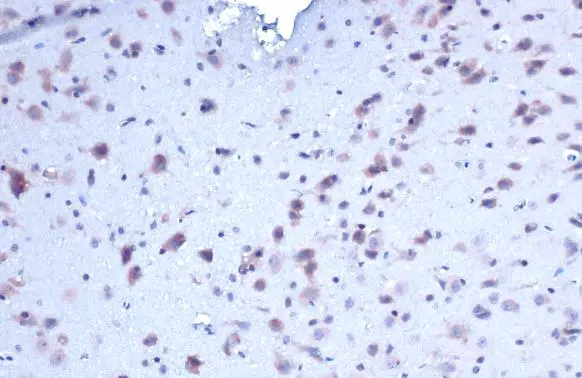
PACAP antibody [N1C3] detects PACAP protein at cytoplasm by immunohistochemical analysis. Sample: Paraffin-embedded rat brain. PACAP stained by PACAP antibody [N1C3] (GTX106794) diluted at 1:200. Antigen Retrieval: Citrate buffer, pH 6.0, 15 min
![PACAP antibody [N1C3] detects PACAP protein at cytoplasm by immunohistochemical analysis. Sample: Paraffin-embedded mouse brain. PACAP stained by PACAP antibody [N1C3] (GTX106794) diluted at 1:200. Antigen Retrieval: Citrate buffer, pH 6.0, 15 min](https://www.genetex.com/upload/website/prouct_img/normal/GTX106794/GTX106794_44713_20220722_IHC-P_M_22081423_872.webp "PACAP antibody [N1C3] detects PACAP protein at cytoplasm by immunohistochemical analysis. Sample: Paraffin-embedded mouse brain. PACAP stained by PACAP antibody [N1C3] (GTX106794) diluted at 1:200. Antigen Retrieval: Citrate buffer, pH 6.0, 15 min")
![PACAP antibody [N1C3] detects PACAP protein at cytoplasm by immunofluorescent analysis. Sample: Raji cells were fixed in 4% paraformaldehyde at RT for 15 min. Green: PACAP stained by PACAP antibody [N1C3] (GTX106794) diluted at 1:500. Blue: Fluoroshield with DAPI (GTX30920).](https://www.genetex.com/upload/website/prouct_img/normal/GTX106794/GTX106794_44713_20220916_ICC_IF_22110201_536.webp "PACAP antibody [N1C3] detects PACAP protein at cytoplasm by immunofluorescent analysis. Sample: Raji cells were fixed in 4% paraformaldehyde at RT for 15 min. Green: PACAP stained by PACAP antibody [N1C3] (GTX106794) diluted at 1:500. Blue: Fluoroshield with DAPI (GTX30920).")
![PACAP antibody [N1C3] detects PACAP protein at cytoplasm in mouse brain by immunohistochemical analysis. Sample:Frozen section of mouse brain. PACAP antibody [N1C3] (GTX106794) diluted at 1:500.](https://www.genetex.com/upload/website/prouct_img/normal/GTX106794/GTX106794_39959_20160126_IHC-Fr_M_w_23060120_801.webp "PACAP antibody [N1C3] detects PACAP protein at cytoplasm in mouse brain by immunohistochemical analysis. Sample:Frozen section of mouse brain. PACAP antibody [N1C3] (GTX106794) diluted at 1:500.")
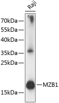
![Mouse tissue extract (50 μg) was separated by 12% SDS-PAGE, and the membrane was blotted with PACAP antibody [N1C3] (GTX106794) diluted at 1:500. The HRP-conjugated anti-rabbit IgG antibody (GTX213110-01) was used to detect the primary antibody, and the signal was developed with Trident ECL plus-Enhanced.](https://www.genetex.com/upload/website/prouct_img/normal/GTX106794/GTX106794_39959_20180907_WB_M_spleen_w_23060120_988.webp "Mouse tissue extract (50 μg) was separated by 12% SDS-PAGE, and the membrane was blotted with PACAP antibody [N1C3] (GTX106794) diluted at 1:500. The HRP-conjugated anti-rabbit IgG antibody (GTX213110-01) was used to detect the primary antibody, and the signal was developed with Trident ECL plus-Enhanced.")
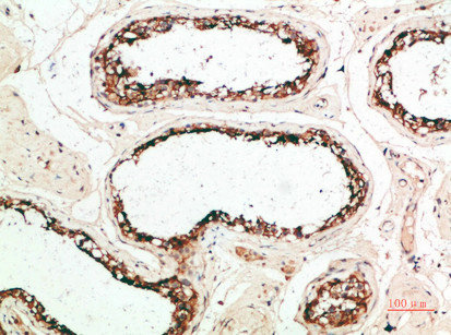
![PACAP antibody [N1C3] detects PACAP protein at cytosol on human normal colon mucosa with lymphocyte by immunohistochemical analysis. Sample: Paraffin-embedded normal colon mucosa with lymphocyte. PACAP antibody [N1C3] (GTX106794) dilution: 1:500.](https://www.genetex.com/upload/website/prouct_img/normal/GTX106794/GTX106794_39959_IHC_w_23060120_538.webp "PACAP antibody [N1C3] detects PACAP protein at cytosol on human normal colon mucosa with lymphocyte by immunohistochemical analysis. Sample: Paraffin-embedded normal colon mucosa with lymphocyte. PACAP antibody [N1C3] (GTX106794) dilution: 1:500.")
![Various whole cell extracts (30 μg) were separated by 15% SDS-PAGE, and the membrane was blotted with PACAP antibody [N1C3] (GTX106794) diluted at 1:1000. The HRP-conjugated anti-rabbit IgG antibody (GTX213110-01) was used to detect the primary antibody.](https://www.genetex.com/upload/website/prouct_img/normal/GTX106794/GTX106794_44713_20220701_WB_24060619_717.webp "Various whole cell extracts (30 μg) were separated by 15% SDS-PAGE, and the membrane was blotted with PACAP antibody [N1C3] (GTX106794) diluted at 1:1000. The HRP-conjugated anti-rabbit IgG antibody (GTX213110-01) was used to detect the primary antibody.")
PACAP antibody [N1C3] detects PACAP protein at cytoplasm by immunohistochemical analysis. Sample: Paraffin-embedded rat brain. PACAP stained by PACAP antibody [N1C3] (GTX106794) diluted at 1:200. Antigen Retrieval: Citrate buffer, pH 6.0, 15 min
PACAP antibody [N1C3]
GTX106794
ApplicationsImmunoFluorescence, Western Blot, ImmunoCytoChemistry, ImmunoHistoChemistry, ImmunoHistoChemistry Frozen, ImmunoHistoChemistry Paraffin
Product group Antibodies
ReactivityHuman, Mouse, Rat
TargetMZB1
Overview
- SupplierGeneTex
- Product NamePACAP antibody [N1C3]
- Delivery Days Customer9
- Application Supplier NoteWB: 1:500-1:3000. IHC-P: 1:100-1:1000. IHC-Fr: 1:100-1:1000. *Optimal dilutions/concentrations should be determined by the researcher.Not tested in other applications.
- ApplicationsImmunoFluorescence, Western Blot, ImmunoCytoChemistry, ImmunoHistoChemistry, ImmunoHistoChemistry Frozen, ImmunoHistoChemistry Paraffin
- CertificationResearch Use Only
- ClonalityPolyclonal
- Concentration0.48 mg/ml
- ConjugateUnconjugated
- Gene ID51237
- Target nameMZB1
- Target descriptionmarginal zone B and B1 cell specific protein
- Target synonymsMEDA-7, PACAP, pERp1, marginal zone B- and B1-cell-specific protein, HSPC190, caspase-2 binding protein, mesenteric estrogen-dependent adipose 7, mesenteric oestrogen-dependent adipose gene- 7, plasma cell-induced ER protein 1, plasma cell-induced resident ER protein, plasma cell-induced resident endoplasmic reticulum protein, proapoptotic caspase adapter protein, proapoptotic caspase adaptor protein
- HostRabbit
- IsotypeIgG
- Protein IDQ8WU39
- Protein NameMarginal zone B- and B1-cell-specific protein
- Scientific DescriptionAssociates with immunoglobulin M (IgM) heavy and light chains and promotes IgM assembly and secretion. May exert its effect by acting as a molecular chaperone or as an oxidoreductase as it displays a low level of oxidoreductase activity (By similarity). Isoform 2 may be involved in regulation of apoptosis.
- ReactivityHuman, Mouse, Rat
- Storage Instruction-20°C or -80°C,2°C to 8°C
- UNSPSC41116161
Datasheet
Related products
Product group Antibodies
Anti-PACAP AntibodyA88543
ApplicationsImmunoFluorescence, Western Blot, ImmunoCytoChemistry, ImmunoHistoChemistry
ReactivityHuman, Mouse, Rat
- SizePrice

Product group Antibodies
Anti-MZB1 Antibody Picoband(r)A08281-1-CARRIER-FREE
ApplicationsFlow Cytometry, ImmunoFluorescence, Western Blot, ELISA, ImmunoCytoChemistry, ImmunoHistoChemistry
ReactivityHuman
TargetMZB1
- SizePrice
Product group Antibodies
Anti-MZB1 Antibody144-63304
ApplicationsImmunoFluorescence, Western Blot, ImmunoHistoChemistry
ReactivityHuman, Mouse, Rat
TargetMZB1
- SizePrice
Product group Antibodies

ApplicationsELISA
ReactivityHuman
TargetMZB1
- SizePrice
Product group Antibodies
MZB1 AntibodyCSB-PA893286
ApplicationsELISA, ImmunoHistoChemistry
ReactivityHuman
TargetMZB1
- SizePrice

Product group Antibodies
Anti-MZB1-25ulHPA043745
ApplicationsWestern Blot, ImmunoHistoChemistry
ReactivityHuman
- SizePrice