


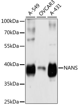
Figure 1. Western blot analysis of NANS using anti-NANS antibody (A08540-2). Electrophoresis was performed on a 5-20% SDS-PAGE gel at 70V (Stacking gel) / 90V (Resolving gel) for 2-3 hours. The sample well of each lane was loaded with 30 ug of sample under reducing conditions. Lane 1: human Hela whole cell lysates, Lane 2: human HepG2 whole cell lysates, Lane 3: human MCF-7 whole cell lysates, Lane 4: rat brain tissue lysates, Lane 5: rat liver tissue lysates. After electrophoresis, proteins were transferred to a nitrocellulose membrane at 150 mA for 50-90 minutes. Blocked the membrane with 5% non-fat milk/TBS for 1.5 hour at RT. The membrane was incubated with rabbit anti-NANS antigen affinity purified polyclonal antibody (A08540-2) at 0.5 microg/mL overnight at 4°C, then washed with TBS-0.1%Tween 3 times with 5 minutes each and probed with a goat anti-rabbit IgG-HRP secondary antibody at a dilution of 1:5000 for 1.5 hour at RT. The signal is developed using an Enhanced Chemiluminescent detection (ECL) kit (Catalog # EK1002) with Tanon 5200 system. A specific band was detected for NANS at approximately 38 kDa. The expected band size for NANS is at 40 kDa.
. NANS was detected in an immunocytochemical section of Hela cells. Enzyme antigen retrieval was performed using IHC enzyme antigen retrieval reagent (AR0022) for 15 mins. The cells were blocked with 10% goat serum. And then incubated with 4 microg/mL rabbit anti-NANS Antibody (A08540-2) overnight at 4°C. DyLight®488 Conjugated Goat Anti-Rabbit IgG (BA1127) was used as secondary antibody at 1:500 dilution and incubated for 30 minutes at 37°C. The section was counterstained with DAPI. Visualize using a fluorescence microscope and filter sets appropriate for the label used.")
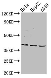
Figure 1. Western blot analysis of NANS using anti-NANS antibody (A08540-2). Electrophoresis was performed on a 5-20% SDS-PAGE gel at 70V (Stacking gel) / 90V (Resolving gel) for 2-3 hours. The sample well of each lane was loaded with 30 ug of sample under reducing conditions. Lane 1: human Hela whole cell lysates, Lane 2: human HepG2 whole cell lysates, Lane 3: human MCF-7 whole cell lysates, Lane 4: rat brain tissue lysates, Lane 5: rat liver tissue lysates. After electrophoresis, proteins were transferred to a nitrocellulose membrane at 150 mA for 50-90 minutes. Blocked the membrane with 5% non-fat milk/TBS for 1.5 hour at RT. The membrane was incubated with rabbit anti-NANS antigen affinity purified polyclonal antibody (A08540-2) at 0.5 microg/mL overnight at 4°C, then washed with TBS-0.1%Tween 3 times with 5 minutes each and probed with a goat anti-rabbit IgG-HRP secondary antibody at a dilution of 1:5000 for 1.5 hour at RT. The signal is developed using an Enhanced Chemiluminescent detection (ECL) kit (Catalog # EK1002) with Tanon 5200 system. A specific band was detected for NANS at approximately 38 kDa. The expected band size for NANS is at 40 kDa.
Anti-NANS Antibody Picoband(r)
A08540-2-CARRIER-FREE
ApplicationsImmunoFluorescence, Western Blot, ELISA, ImmunoCytoChemistry
Product group Antibodies
ReactivityHuman, Rat
TargetNANS
Overview
- SupplierBoster Bio
- Product NameAnti-NANS Antibody Picoband(r)
- Delivery Days Customer9
- ApplicationsImmunoFluorescence, Western Blot, ELISA, ImmunoCytoChemistry
- CertificationResearch Use Only
- ClonalityPolyclonal
- Concentration500 ug/ml
- Gene ID54187
- Target nameNANS
- Target descriptionN-acetylneuraminate synthase
- Target synonymsHEL-S-100, SAS, SEMDCG, SEMDG, N-acetylneuraminate-9-phosphate synthase, 3-deoxy-D-glycero-D-galacto-nononate 9-phosphate synthase, N-acetylneuraminic acid phosphate synthase, N-acetylneuraminic acid synthase, epididymis secretory protein Li 100, sialic acid phosphate synthase, sialic acid synthase
- HostRabbit
- IsotypeIgG
- Protein IDQ9NR45
- Protein NameN-acetylneuraminate-9-phosphate synthase
- Scientific DescriptionBoster Bio Anti-NANS Antibody Picoband® catalog # A08540-2. Tested in WB, ICC/IF, ELISA applications. This antibody reacts with Human, Rat. The brand Picoband indicates this is a premium antibody that guarantees superior quality, high affinity, and strong signals with minimal background in Western blot applications. Only our best-performing antibodies are designated as Picoband, ensuring unmatched performance.
- ReactivityHuman, Rat
- Storage Instruction-20°C,2°C to 8°C
- UNSPSC12352203
Related products
Product group Antibodies
Anti-NANS Antibody144-61396
ApplicationsWestern Blot
ReactivityHuman
TargetNANS
- SizePrice

Product group Antibodies
NANS AntibodyCSB-PA885699LA01HU
ApplicationsImmunoFluorescence, Western Blot, ELISA, ImmunoHistoChemistry
ReactivityHuman
TargetNANS
- SizePrice
Product group Antibodies
NANS Polyclonal AntibodyCAC13524
ApplicationsImmunoFluorescence, Western Blot, ELISA, ImmunoHistoChemistry
TargetNANS
- SizePrice
Product group Antibodies
NANS antibody [N1C3]GTX105223
ApplicationsImmunoFluorescence, Western Blot, ImmunoCytoChemistry, ImmunoHistoChemistry, ImmunoHistoChemistry Paraffin
ReactivityHuman
TargetNANS
- SizePrice
Product group Antibodies
Anti-NANS AntibodyHPA019223
ApplicationsWestern Blot, ImmunoCytoChemistry, ImmunoHistoChemistry
ReactivityHuman, Mouse, Rat
TargetNANS
- SizePrice
Product group Antibodies
Anti-NANS AntibodyCAB4778
ApplicationsWestern Blot, ELISA
ReactivityHuman
TargetNANS
- SizePrice