


Figure 1. Western blot analysis of NDUFAB1 using anti-NDUFAB1 antibody (M09620). Electrophoresis was performed on a 5-20% SDS-PAGE gel at 70V (Stacking gel) / 90V (Resolving gel) for 2-3 hours. The sample well of each lane was loaded with 30 ug of sample under reducing conditions. Lane 1: human A431 whole cell lysates, Lane 2: human HepG2 whole cell lysates, Lane 3: human MCF-7 whole cell lysates, Lane 4: human 293T whole cell lysates, Lane 5: human THP-1 whole cell lysates, Lane 6: human PC-3 whole cell lysates, Lane 7: rat RH35 whole cell lysates, Lane 8: mouse NIH/3T3 whole cell lysates. After electrophoresis, proteins were transferred to a nitrocellulose membrane at 150 mA for 50-90 minutes. Blocked the membrane with 5% non-fat milk/TBS for 1.5 hour at RT. The membrane was incubated with rabbit anti-NDUFAB1 antigen affinity purified monoclonal antibody (Catalog # M09620) at 1:500 overnight at 4°C, then washed with TBS-0.1%Tween 3 times with 5 minutes each and probed with a goat anti-rabbit IgG-HRP secondary antibody at a dilution of 1:500 for 1.5 hour at RT. The signal is developed using an Enhanced Chemiluminescent detection (ECL) kit (Catalog # EK1002) with Tanon 5200 system. A specific band was detected for NDUFAB1 at approximately 14 kDa. The expected band size for NDUFAB1 is at 17 kDa.
. NDUFAB1 was detected in a paraffin-embedded section of human colorectal adenocarcinoma tissue. Heat mediated antigen retrieval was performed in EDTA buffer (pH 8.0, epitope retrieval solution). The tissue section was blocked with 10% goat serum. The tissue section was then incubated with 1:50 rabbit anti-NDUFAB1 Antibody (M09620) overnight at 4°C. Peroxidase Conjugated Goat Anti-rabbit IgG was used as secondary antibody and incubated for 30 minutes at 37°C. The tissue section was developed using HRP Conjugated Rabbit IgG Super Vision Assay Kit (Catalog # SV0002) with DAB as the chromogen.")
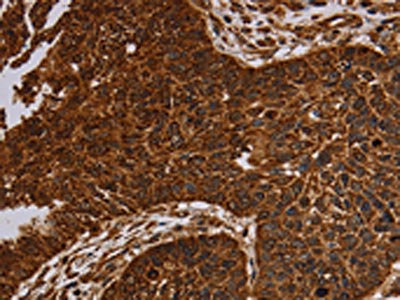
. NDUFAB1 was detected in a paraffin-embedded section of human thyroid cancer tissue. Heat mediated antigen retrieval was performed in EDTA buffer (pH 8.0, epitope retrieval solution). The tissue section was blocked with 10% goat serum. The tissue section was then incubated with 1:50 rabbit anti-NDUFAB1 Antibody (M09620) overnight at 4°C. Peroxidase Conjugated Goat Anti-rabbit IgG was used as secondary antibody and incubated for 30 minutes at 37°C. The tissue section was developed using HRP Conjugated Rabbit IgG Super Vision Assay Kit (Catalog # SV0002) with DAB as the chromogen.")
. NDUFAB1 was detected in a paraffin-embedded section of human breast cancer tissue. Heat mediated antigen retrieval was performed in EDTA buffer (pH 8.0, epitope retrieval solution). The tissue section was blocked with 10% goat serum. The tissue section was then incubated with 1:50 rabbit anti-NDUFAB1 Antibody (M09620) overnight at 4°C. Peroxidase Conjugated Goat Anti-rabbit IgG was used as secondary antibody and incubated for 30 minutes at 37°C. The tissue section was developed using HRP Conjugated Rabbit IgG Super Vision Assay Kit (Catalog # SV0002) with DAB as the chromogen.")
. NDUFAB1 was detected in a paraffin-embedded section of human placenta tissue. Heat mediated antigen retrieval was performed in EDTA buffer (pH 8.0, epitope retrieval solution). The tissue section was blocked with 10% goat serum. The tissue section was then incubated with 1:50 rabbit anti-NDUFAB1 Antibody (M09620) overnight at 4°C. Peroxidase Conjugated Goat Anti-rabbit IgG was used as secondary antibody and incubated for 30 minutes at 37°C. The tissue section was developed using HRP Conjugated Rabbit IgG Super Vision Assay Kit (Catalog # SV0002) with DAB as the chromogen.")
. NDUFAB1 was detected in a paraffin-embedded section of human testicular germ cell tumor tissue. Heat mediated antigen retrieval was performed in EDTA buffer (pH 8.0, epitope retrieval solution). The tissue section was blocked with 10% goat serum. The tissue section was then incubated with 1:50 rabbit anti-NDUFAB1 Antibody (M09620) overnight at 4°C. Peroxidase Conjugated Goat Anti-rabbit IgG was used as secondary antibody and incubated for 30 minutes at 37°C. The tissue section was developed using HRP Conjugated Rabbit IgG Super Vision Assay Kit (Catalog # SV0002) with DAB as the chromogen.")
Figure 1. Western blot analysis of NDUFAB1 using anti-NDUFAB1 antibody (M09620). Electrophoresis was performed on a 5-20% SDS-PAGE gel at 70V (Stacking gel) / 90V (Resolving gel) for 2-3 hours. The sample well of each lane was loaded with 30 ug of sample under reducing conditions. Lane 1: human A431 whole cell lysates, Lane 2: human HepG2 whole cell lysates, Lane 3: human MCF-7 whole cell lysates, Lane 4: human 293T whole cell lysates, Lane 5: human THP-1 whole cell lysates, Lane 6: human PC-3 whole cell lysates, Lane 7: rat RH35 whole cell lysates, Lane 8: mouse NIH/3T3 whole cell lysates. After electrophoresis, proteins were transferred to a nitrocellulose membrane at 150 mA for 50-90 minutes. Blocked the membrane with 5% non-fat milk/TBS for 1.5 hour at RT. The membrane was incubated with rabbit anti-NDUFAB1 antigen affinity purified monoclonal antibody (Catalog # M09620) at 1:500 overnight at 4°C, then washed with TBS-0.1%Tween 3 times with 5 minutes each and probed with a goat anti-rabbit IgG-HRP secondary antibody at a dilution of 1:500 for 1.5 hour at RT. The signal is developed using an Enhanced Chemiluminescent detection (ECL) kit (Catalog # EK1002) with Tanon 5200 system. A specific band was detected for NDUFAB1 at approximately 14 kDa. The expected band size for NDUFAB1 is at 17 kDa.
Anti-NDUFAB1 Rabbit Monoclonal Antibody
M09620
ApplicationsFlow Cytometry, ImmunoPrecipitation, Western Blot, ImmunoHistoChemistry
Product group Antibodies
ReactivityHuman, Mouse, Rat
TargetNDUFAB1
Overview
- SupplierBoster Bio
- Product NameAnti-NDUFAB1 Rabbit Monoclonal Antibody
- Delivery Days Customer9
- ApplicationsFlow Cytometry, ImmunoPrecipitation, Western Blot, ImmunoHistoChemistry
- CertificationResearch Use Only
- ClonalityMonoclonal
- Clone ID30N94
- Gene ID4706
- Target nameNDUFAB1
- Target descriptionNADH:ubiquinone oxidoreductase subunit AB1
- Target synonymsACP, ACP1, FASN2A, SDAP, acyl carrier protein, mitochondrial, CI-SDAP, NADH dehydrogenase (ubiquinone) 1, alpha/beta subcomplex, 1, 8kDa, NADH-ubiquinone oxidoreductase 9.6 kDa subunit, NADH:ubiquinone oxidoreductase SDAP subunit, complex I SDAP subunit, mitochondrial acyl carrier protein
- HostRabbit
- IsotypeIgG
- Protein IDO14561
- Protein NameAcyl carrier protein, mitochondrial
- Scientific DescriptionBoster Bio Anti-NDUFAB1 Rabbit Monoclonal Antibody catalog # M09620. Tested in WB, IHC, IP, Flow Cytometry applications. This antibody reacts with Human, Mouse, Rat.
- ReactivityHuman, Mouse, Rat
- Storage Instruction-20°C
- UNSPSC12352203
Related products
Product group Antibodies
ApplicationsImmunoPrecipitation, Western Blot, ImmunoCytoChemistry, ImmunoHistoChemistry
ReactivityMouse
TargetNDUFAB1
- SizePrice
Product group Antibodies
NDUFAB1 AntibodyCSB-PA446344
ApplicationsELISA, ImmunoHistoChemistry
ReactivityHuman, Mouse
TargetNDUFAB1
- SizePrice
Product group Antibodies
Anti-NDUFAB1 Antibody144-60741
ApplicationsWestern Blot
ReactivityHuman, Mouse
TargetNDUFAB1
- SizePrice

Product group Antibodies
Anti-NDUFAB1 AntibodyA88206
ApplicationsWestern Blot
ReactivityHuman, Mouse, Rat
- SizePrice

Product group Antibodies
Anti-NDUFAB1 AntibodyHPA054364
ApplicationsImmunoCytoChemistry, ImmunoHistoChemistry
ReactivityHuman
TargetNDUFAB1
- SizePrice
Product group Antibodies

NDUFAB1 / ACP AntibodyLS-C403360
ApplicationsELISA, ImmunoHistoChemistry
ReactivityHuman, Mouse
TargetNDUFAB1
- SizePrice
Product group Antibodies
NDUFAB1 Recombinant AntibodyBSM-62777R
ApplicationsFlow Cytometry, ImmunoFluorescence, ImmunoPrecipitation, Western Blot, ImmunoHistoChemistry, ImmunoHistoChemistry Frozen, ImmunoHistoChemistry Paraffin
ReactivityHuman
TargetNDUFAB1
- SizePrice