


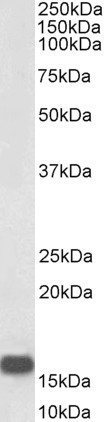
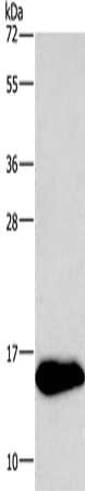
Figure 1. Western blot analysis of Neurogranin/NRGN using anti-Neurogranin/NRGN antibody (A05781-3). Electrophoresis was performed on a 5-20% SDS-PAGE gel at 70V (Stacking gel) / 90V (Resolving gel) for 2-3 hours. The sample well of each lane was loaded with 30 ug of sample under reducing conditions. Lane 1: human PC-3 whole cell lysates, Lane 2: rat brain tissue lysates, Lane 3: mouse brain tissue lysates. After electrophoresis, proteins were transferred to a nitrocellulose membrane at 150 mA for 50-90 minutes. Blocked the membrane with 5% non-fat milk/TBS for 1.5 hour at RT. The membrane was incubated with rabbit anti-Neurogranin/NRGN antigen affinity purified polyclonal antibody (Catalog # A05781-3) at 0.25 microg/mL overnight at 4°C, then washed with TBS-0.1%Tween 3 times with 5 minutes each and probed with a goat anti-rabbit IgG-HRP secondary antibody at a dilution of 1:5000 for 1.5 hour at RT. The signal is developed using an Enhanced Chemiluminescent detection (ECL) kit (Catalog # EK1002) with Tanon 5200 system. A specific band was detected for Neurogranin/NRGN at approximately 11 kDa. The expected band size for Neurogranin/NRGN is at 8 kDa.
. Overlay histogram showing K562 cells stained with A05781-3 (Blue line). To facilitate intracellular staining, cells were fixed with 4% paraformaldehyde and permeabilized with permeabilization buffer. The cells were blocked with 10% normal goat serum. And then incubated with rabbit anti-Neurogranin/NRGN Antibody (A05781-3, 1 microg/1x106 cells) for 30 min at 20°C. DyLight®488 conjugated goat anti-rabbit IgG (BA1127, 5-10 microg/1x106 cells) was used as secondary antibody for 30 minutes at 20°C. Isotype control antibody (Green line) was rabbit IgG (1 microg/1x106) used under the same conditions. Unlabelled sample without incubation with primary antibody and secondary antibody (Red line) was used as a blank control.")
Figure 1. Western blot analysis of Neurogranin/NRGN using anti-Neurogranin/NRGN antibody (A05781-3). Electrophoresis was performed on a 5-20% SDS-PAGE gel at 70V (Stacking gel) / 90V (Resolving gel) for 2-3 hours. The sample well of each lane was loaded with 30 ug of sample under reducing conditions. Lane 1: human PC-3 whole cell lysates, Lane 2: rat brain tissue lysates, Lane 3: mouse brain tissue lysates. After electrophoresis, proteins were transferred to a nitrocellulose membrane at 150 mA for 50-90 minutes. Blocked the membrane with 5% non-fat milk/TBS for 1.5 hour at RT. The membrane was incubated with rabbit anti-Neurogranin/NRGN antigen affinity purified polyclonal antibody (Catalog # A05781-3) at 0.25 microg/mL overnight at 4°C, then washed with TBS-0.1%Tween 3 times with 5 minutes each and probed with a goat anti-rabbit IgG-HRP secondary antibody at a dilution of 1:5000 for 1.5 hour at RT. The signal is developed using an Enhanced Chemiluminescent detection (ECL) kit (Catalog # EK1002) with Tanon 5200 system. A specific band was detected for Neurogranin/NRGN at approximately 11 kDa. The expected band size for Neurogranin/NRGN is at 8 kDa.
Anti-Neurogranin/NRGN Antibody Picoband(r)
A05781-3-CARRIER-FREE
ApplicationsFlow Cytometry, Western Blot, ELISA
Product group Antibodies
ReactivityHuman, Mouse, Rat
TargetNRGN
Overview
- SupplierBoster Bio
- Product NameAnti-Neurogranin/NRGN Antibody Picoband(r)
- Delivery Days Customer9
- ApplicationsFlow Cytometry, Western Blot, ELISA
- CertificationResearch Use Only
- ClonalityPolyclonal
- Concentration500 ug/ml
- Gene ID4900
- Target nameNRGN
- Target descriptionneurogranin
- Target synonymsRC3, hng, neurogranin, calmodulin-binding protein, neurogranin (protein kinase C substrate, RC3), protein kinase C substrate
- HostRabbit
- IsotypeIgG
- Protein IDQ92686
- Protein NameNeurogranin
- Scientific DescriptionBoster Bio Anti-Neurogranin/NRGN Antibody Picoband® catalog # A05781-3. Tested in ELISA, Flow Cytometry, IHC-P, WB applications. This antibody reacts with Human, Mouse, Rat. The brand Picoband indicates this is a premium antibody that guarantees superior quality, high affinity, and strong signals with minimal background in Western blot applications. Only our best-performing antibodies are designated as Picoband, ensuring unmatched performance.
- ReactivityHuman, Mouse, Rat
- Storage Instruction-20°C,2°C to 8°C
- UNSPSC12352203
Related products
Product group Antibodies
ApplicationsWestern Blot, ELISA
ReactivityHuman
- SizePrice
Product group Antibodies
Anti-NRGN Antibody144-08444
ApplicationsWestern Blot
ReactivityHuman, Mouse, Rat
TargetNRGN
- SizePrice
Product group Antibodies
Neurogranin Polyclonal AntibodyBS-11435R
ApplicationsImmunoFluorescence, Western Blot, ELISA, ImmunoCytoChemistry, ImmunoHistoChemistry, ImmunoHistoChemistry Frozen, ImmunoHistoChemistry Paraffin
ReactivityBovine, Canine, Equine, Human, Mouse, Porcine, Rabbit, Rat
TargetNRGN
- SizePrice
Product group Antibodies

ApplicationsWestern Blot, ELISA
ReactivityBovine, Canine, Human, Mouse
TargetNRGN
- SizePrice
Product group Antibodies
NRGN AntibodyCSB-PA793948
ApplicationsWestern Blot, ELISA
ReactivityHuman, Mouse, Rat
TargetNRGN
- SizePrice
Product group Antibodies
ApplicationsFlow Cytometry
TargetNRGN
- SizePrice
Product group Antibodies

NRGN / Neurogranin AntibodyLS-C409979
ApplicationsWestern Blot
ReactivityHuman, Mouse, Rat
TargetNRGN
- SizePrice
![Neurogranin antibody detects Neurogranin protein by immunofluorescent analysis. Sample: DIV9 rat E18 primary hippocampal neuron cells were fixed in 4% paraformaldehyde at RT for 15 min. Green: Neurogranin stained by Neurogranin antibody (GTX134048) diluted at 1:500. Red: beta Tubulin 3/ Tuj1, stained by beta Tubulin 3/ Tuj1 antibody [GT11710] (GTX631836) diluted at 1:500. Blue: Fluoroshield with DAPI (GTX30920).](https://www.genetex.com/upload/website/prouct_img/normal/GTX134048/GTX134048_43075_20180613_ICC_IF_R_w_23060620_398.webp)
Product group Antibodies
Neurogranin antibodyGTX134048
ApplicationsImmunoFluorescence, Western Blot, ImmunoCytoChemistry
ReactivityHuman, Mouse, Rat
TargetNRGN
- SizePrice