


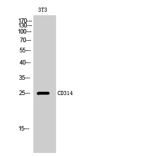
Figure 1. Western blot analysis of NKG2D using anti-NKG2D antibody (A00661-1). Electrophoresis was performed on a 5-20% SDS-PAGE gel at 70V (Stacking gel) / 90V (Resolving gel) for 2-3 hours. The sample well of each lane was loaded with 50ug of sample under reducing conditions. Lane 1: rat lymph tissue lysates, Lane 2: rat spleen tissue lysates, Lane 3: mouse thymus tissue lysates. After Electrophoresis, proteins were transferred to a Nitrocellulose membrane at 150mA for 50-90 minutes. Blocked the membrane with 5% Non-fat Milk/ TBS for 1.5 hour at RT. The membrane was incubated with rabbit anti-NKG2D antigen affinity purified polyclonal antibody (Catalog # A00661-1) at 0.5 microg/mL overnight at 4°C, then washed with TBS-0.1%Tween 3 times with 5 minutes each and probed with a goat anti-rabbit IgG-HRP secondary antibody at a dilution of 1:10000 for 1.5 hour at RT. The signal is developed using an Enhanced Chemiluminescent detection (ECL) kit (Catalog # EK1002) with Tanon 5200 system. A specific band was detected for NKG2D at approximately 35KD. The expected band size for NKG2D is at 25KD.
Figure 1. Western blot analysis of NKG2D using anti-NKG2D antibody (A00661-1). Electrophoresis was performed on a 5-20% SDS-PAGE gel at 70V (Stacking gel) / 90V (Resolving gel) for 2-3 hours. The sample well of each lane was loaded with 50ug of sample under reducing conditions. Lane 1: rat lymph tissue lysates, Lane 2: rat spleen tissue lysates, Lane 3: mouse thymus tissue lysates. After Electrophoresis, proteins were transferred to a Nitrocellulose membrane at 150mA for 50-90 minutes. Blocked the membrane with 5% Non-fat Milk/ TBS for 1.5 hour at RT. The membrane was incubated with rabbit anti-NKG2D antigen affinity purified polyclonal antibody (Catalog # A00661-1) at 0.5 microg/mL overnight at 4°C, then washed with TBS-0.1%Tween 3 times with 5 minutes each and probed with a goat anti-rabbit IgG-HRP secondary antibody at a dilution of 1:10000 for 1.5 hour at RT. The signal is developed using an Enhanced Chemiluminescent detection (ECL) kit (Catalog # EK1002) with Tanon 5200 system. A specific band was detected for NKG2D at approximately 35KD. The expected band size for NKG2D is at 25KD.
Anti-NKG2D/KLRK1 Antibody Picoband(r)
A00661-1-CARRIER-FREE
ApplicationsWestern Blot
Product group Antibodies
ReactivityHuman, Mouse, Rat
TargetKLRK1
Overview
- SupplierBoster Bio
- Product NameAnti-NKG2D/KLRK1 Antibody Picoband(r)
- Delivery Days Customer9
- ApplicationsWestern Blot
- CertificationResearch Use Only
- ClonalityPolyclonal
- Concentration500 ug/ml
- Gene ID22914
- Target nameKLRK1
- Target descriptionkiller cell lectin like receptor K1
- Target synonymsCD314, D12S2489E, KLR, NKG2-D, NKG2D, NKG2-D type II integral membrane protein, NK cell receptor D, NKG2-D-activating NK receptor, killer cell lectin-like receptor subfamily K, member 1
- HostRabbit
- IsotypeIgG
- Protein IDP26718
- Protein NameNKG2-D type II integral membrane protein
- Scientific DescriptionBoster Bio Anti-NKG2D/KLRK1 Antibody Picoband® catalog # A00661-1. Tested in WB applications. This antibody reacts with Mouse, Rat. The brand Picoband indicates this is a premium antibody that guarantees superior quality, high affinity, and strong signals with minimal background in Western blot applications. Only our best-performing antibodies are designated as Picoband, ensuring unmatched performance.
- ReactivityHuman, Mouse, Rat
- Storage Instruction-20°C,2°C to 8°C
- UNSPSC12352203
Related products
Product group Antibodies

Anti-NKG2D [6E5A7]Ab02029-10.0
ApplicationsFlow Cytometry, Neutralisation/Blocking
ReactivityHuman
TargetKLRK1
- SizePrice
Product group Antibodies
Anti-KLRK1 AntibodyA101001
ApplicationsWestern Blot, ELISA
ReactivityHuman
- SizePrice
Product group Antibodies
Anti-KLRK1 Antibody144-66050
ApplicationsWestern Blot
ReactivityHuman, Mouse
TargetKLRK1
- SizePrice
Product group Antibodies
KLRK1 AntibodyCSB-PA008277
ApplicationsWestern Blot, ELISA
ReactivityHuman
TargetKLRK1
- SizePrice
Product group Antibodies

Goat anti-NKG2D / KLRK1EB06839
ApplicationsFlow Cytometry, Western Blot, ELISA
ReactivityHuman, Mouse, Rat
TargetKLRK1
- SizePrice
Product group Antibodies
Klrk1 Polyclonal AntibodyCAC08581
ApplicationsImmunoFluorescence, Western Blot, ELISA, ImmunoHistoChemistry
TargetKLRK1
- SizePrice
Product group Antibodies

KLRK1 / CD314 / NKG2D AntibodyLS-C335641
ApplicationsWestern Blot
ReactivityHuman, Mouse
TargetKLRK1
- SizePrice
![NKG2D antibody [N3C2], Internal detects NKG2D protein at cell membrane by immunofluorescent analysis. Sample: HepG2 cells were fixed in 4% paraformaldehyde at RT for 15 min. Green: NKG2D stained by NKG2D antibody [N3C2], Internal (GTX104761) diluted at 1:500. Blue: Fluoroshield with DAPI (GTX30920). Scale bar= 10μm.](https://www.genetex.com/upload/website/prouct_img/normal/GTX104761/GTX104761_44510_20230210_ICC_IF_23021401_259.webp)
Product group Antibodies
NKG2D antibody [N3C2], InternalGTX104761
ApplicationsImmunoFluorescence, Western Blot, ImmunoCytoChemistry, ImmunoHistoChemistry, ImmunoHistoChemistry Paraffin
ReactivityHuman
TargetKLRK1
- SizePrice