


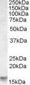
Figure 1. Western blot analysis of PMAIP1 using anti-PMAIP1 antibody (M02287). Electrophoresis was performed on a 5-20% SDS-PAGE gel at 70V (Stacking gel) / 90V (Resolving gel) for 2-3 hours. The sample well of each lane was loaded with 30 ug of sample under reducing conditions. Lane 1: human Jurkat whole cell lysates, Lane 2: human 293T whole cell lysates, Lane 3: rat PC-12 whole cell lysates, Lane 4: mouse RAW264.7 whole cell lysates. After electrophoresis, proteins were transferred to a nitrocellulose membrane at 150 mA for 50-90 minutes. Blocked the membrane with 5% non-fat milk/TBS for 1.5 hour at RT. The membrane was incubated with rabbit anti-PMAIP1 antigen affinity purified monoclonal antibody (Catalog # M02287) at 1:500 overnight at 4°C, then washed with TBS-0.1%Tween 3 times with 5 minutes each and probed with a goat anti-rabbit IgG-HRP secondary antibody at a dilution of 1:500 for 1.5 hour at RT. The signal is developed using an Enhanced Chemiluminescent detection (ECL) kit (Catalog # EK1002) with Tanon 5200 system. A specific band was detected for PMAIP1 at approximately 16 kDa. The expected band size for PMAIP1 is at 6 kDa.
Figure 1. Western blot analysis of PMAIP1 using anti-PMAIP1 antibody (M02287). Electrophoresis was performed on a 5-20% SDS-PAGE gel at 70V (Stacking gel) / 90V (Resolving gel) for 2-3 hours. The sample well of each lane was loaded with 30 ug of sample under reducing conditions. Lane 1: human Jurkat whole cell lysates, Lane 2: human 293T whole cell lysates, Lane 3: rat PC-12 whole cell lysates, Lane 4: mouse RAW264.7 whole cell lysates. After electrophoresis, proteins were transferred to a nitrocellulose membrane at 150 mA for 50-90 minutes. Blocked the membrane with 5% non-fat milk/TBS for 1.5 hour at RT. The membrane was incubated with rabbit anti-PMAIP1 antigen affinity purified monoclonal antibody (Catalog # M02287) at 1:500 overnight at 4°C, then washed with TBS-0.1%Tween 3 times with 5 minutes each and probed with a goat anti-rabbit IgG-HRP secondary antibody at a dilution of 1:500 for 1.5 hour at RT. The signal is developed using an Enhanced Chemiluminescent detection (ECL) kit (Catalog # EK1002) with Tanon 5200 system. A specific band was detected for PMAIP1 at approximately 16 kDa. The expected band size for PMAIP1 is at 6 kDa.
Anti-Noxa Rabbit Monoclonal Antibody
M02287
ApplicationsWestern Blot
Product group Antibodies
ReactivityHuman, Mouse, Rat
TargetPMAIP1
Overview
- SupplierBoster Bio
- Product NameAnti-Noxa Rabbit Monoclonal Antibody
- Delivery Days Customer9
- ApplicationsWestern Blot
- CertificationResearch Use Only
- ClonalityMonoclonal
- Clone IDAAFB-16
- Gene ID5366
- Target namePMAIP1
- Target descriptionphorbol-12-myristate-13-acetate-induced protein 1
- Target synonymsAPR, NOXA, phorbol-12-myristate-13-acetate-induced protein 1, PMA-induced protein 1, adult T cell leukemia-derived PMA-responsive, immediate-early-response protein APR, protein Noxa
- HostRabbit
- IsotypeIgG
- Protein IDQ13794
- Protein NamePhorbol-12-myristate-13-acetate-induced protein 1
- Scientific DescriptionBoster Bio Anti-Noxa Rabbit Monoclonal Antibody catalog # M02287. Tested in WB application. This antibody reacts with Human, Mouse, Rat.
- ReactivityHuman, Mouse, Rat
- Storage Instruction-20°C
- UNSPSC12352203
References
- Fan P, Wang J, Li R, et al. Development and validation of an endoplasmic reticulum stress-related molecular prognostic model for breast cancer. Front Oncol. 2023,13:1178595. doi: 10.3389/fonc.2023.1178595Read this paper
- Chen Y, Yang P, Wang J, et al. p53 directly downregulates the expression of CDC20 to exert anti-tumor activity in mantle cell lymphoma. Exp Hematol Oncol. 2023,12(1):28. doi: 10.1186/s40164-023-00381-7Read this paper
Datasheet
MSDS
Related products
Product group Antibodies
PMAIP1 AntibodyCSB-PA018229LA01HU
ApplicationsImmunoFluorescence, ELISA, ImmunoHistoChemistry
ReactivityHuman
TargetPMAIP1
- SizePrice
Product group Antibodies
Anti-PMAIP1 AntibodyA45471
ApplicationsImmunoHistoChemistry
ReactivityHuman
- SizePrice
Product group Antibodies

Goat anti-NOXAEB09119
ApplicationsWestern Blot, ELISA
ReactivityHuman
TargetPMAIP1
- SizePrice
Product group Antibodies

PMAIP1 / NOXA AntibodyLS-C668202
ApplicationsWestern Blot
ReactivityHuman
TargetPMAIP1
- SizePrice
Product group Antibodies
Noxa antibody, InternalGTX88398
ApplicationsWestern Blot
ReactivityHuman
TargetPMAIP1
- SizePrice
Product group Antibodies
Noxa Recombinant Antibody, AbBy Fluor-350 ConjugatedBSM-61443R-BF350
ApplicationsWestern Blot
ReactivityHuman
TargetPMAIP1
- SizePrice
Product group Antibodies
Anti-PMAIP1 Antibody144-09801
ApplicationsWestern Blot
ReactivityHuman
TargetPMAIP1
- SizePrice