

Anti-PMAIP1 Antibody
144-09801
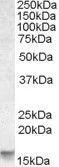
ApplicationsWestern Blot
Product group Antibodies
ReactivityHuman
TargetPMAIP1
Overview
- SupplierRayBiotech
- Product NameAnti-PMAIP1 Antibody
- Delivery Days Customer16
- ApplicationsWestern Blot
- CertificationResearch Use Only
- ClonalityPolyclonal
- ConjugateUnconjugated
- Gene ID5366
- Target namePMAIP1
- Target descriptionphorbol-12-myristate-13-acetate-induced protein 1
- Target synonymsAPR, NOXA, phorbol-12-myristate-13-acetate-induced protein 1, PMA-induced protein 1, adult T cell leukemia-derived PMA-responsive, immediate-early-response protein APR, protein Noxa
- HostRabbit
- IsotypeIgG
- Protein IDQ13794
- Protein NamePhorbol-12-myristate-13-acetate-induced protein 1
- Scientific DescriptionPMAIP1 Polyclonal Antibody
- ReactivityHuman
- Storage Instruction-20°C
- UNSPSC12352203
Related products
Product group Antibodies
PMAIP1 AntibodyCSB-PA018229LA01HU
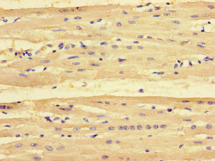
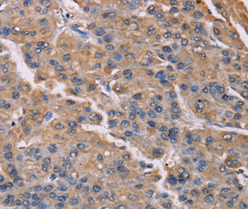
ApplicationsImmunoFluorescence, ELISA, ImmunoHistoChemistry
ReactivityHuman
TargetPMAIP1
- SizePrice
Product group Antibodies
Anti-PMAIP1 AntibodyA45471
ApplicationsImmunoHistoChemistry
ReactivityHuman
- SizePrice

Product group Antibodies
References
ApplicationsWestern Blot
ReactivityHuman, Mouse, Rat
TargetPMAIP1
- SizePrice
Product group Antibodies

Goat anti-NOXAEB09119
ApplicationsWestern Blot, ELISA
ReactivityHuman
TargetPMAIP1
- SizePrice
Product group Antibodies

PMAIP1 / NOXA AntibodyLS-C668202
ApplicationsWestern Blot
ReactivityHuman
TargetPMAIP1
- SizePrice
Product group Antibodies
Noxa antibody, InternalGTX88398
ApplicationsWestern Blot
ReactivityHuman
TargetPMAIP1
- SizePrice
Product group Antibodies
Noxa Recombinant Antibody, AbBy Fluor-350 ConjugatedBSM-61443R-BF350
ApplicationsWestern Blot
ReactivityHuman
TargetPMAIP1
- SizePrice