

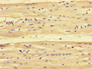
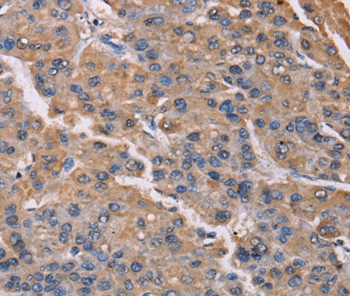
Immunohistochemistry of paraffin-embedded human heart tissue using CSB-PA018229LA01HU at dilution of 1:100

")
Immunohistochemistry of paraffin-embedded human heart tissue using CSB-PA018229LA01HU at dilution of 1:100
PMAIP1 Antibody
CSB-PA018229LA01HU
ApplicationsImmunoFluorescence, ELISA, ImmunoHistoChemistry
Product group Antibodies
ReactivityHuman
TargetPMAIP1
Overview
- SupplierCusabio
- Product NamePMAIP1 Antibody
- Delivery Days Customer20
- ApplicationsImmunoFluorescence, ELISA, ImmunoHistoChemistry
- CertificationResearch Use Only
- ClonalityPolyclonal
- ConjugateUnconjugated
- Gene ID5366
- Target namePMAIP1
- Target descriptionphorbol-12-myristate-13-acetate-induced protein 1
- Target synonymsAPR, NOXA, phorbol-12-myristate-13-acetate-induced protein 1, PMA-induced protein 1, adult T cell leukemia-derived PMA-responsive, immediate-early-response protein APR, protein Noxa
- HostRabbit
- IsotypeIgG
- Protein IDQ13794
- Protein NamePhorbol-12-myristate-13-acetate-induced protein 1
- Scientific DescriptionPromotes activation of caspases and apoptosis. Promotes mitochondrial membrane changes and efflux of apoptogenic proteins from the mitochondria. Contributes to p53/TP53-dependent apoptosis after radiation exposure. Promotes proteasomal degradation of MCL1. Competes with BAK1 for binding to MCL1 and can displace BAK1 from its binding site on MCL1 (By similarity). Competes with BIM/BCL2L11 for binding to MCL1 and can displace BIM/BCL2L11 from its binding site on MCL1.
- ReactivityHuman
- Storage Instruction-20°C or -80°C
- UNSPSC41116161
Related products
Product group Antibodies
Anti-PMAIP1 AntibodyA45471
ApplicationsImmunoHistoChemistry
ReactivityHuman
- SizePrice

Product group Antibodies
References
ApplicationsWestern Blot
ReactivityHuman, Mouse, Rat
TargetPMAIP1
- SizePrice
Product group Antibodies

Goat anti-NOXAEB09119
ApplicationsWestern Blot, ELISA
ReactivityHuman
TargetPMAIP1
- SizePrice
Product group Antibodies

PMAIP1 / NOXA AntibodyLS-C668202
ApplicationsWestern Blot
ReactivityHuman
TargetPMAIP1
- SizePrice
Product group Antibodies
Noxa antibody, InternalGTX88398
ApplicationsWestern Blot
ReactivityHuman
TargetPMAIP1
- SizePrice
Product group Antibodies
Noxa Recombinant Antibody, AbBy Fluor-350 ConjugatedBSM-61443R-BF350
ApplicationsWestern Blot
ReactivityHuman
TargetPMAIP1
- SizePrice
Product group Antibodies
Anti-PMAIP1 Antibody144-09801
ApplicationsWestern Blot
ReactivityHuman
TargetPMAIP1
- SizePrice