


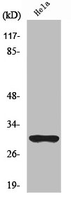
Figure 1. Western blot analysis of NQO1 using anti-NQO1 antibody (A00494-2). Electrophoresis was performed on a 5-20% SDS-PAGE gel at 70V (Stacking gel) / 90V (Resolving gel) for 2-3 hours. The sample well of each lane was loaded with 30 ug of sample under reducing conditions. Lane 1: human MCF-7 whole cell lysates, Lane 2: human Hela whole cell lysates, Lane 3: human A549 whole cell lysates. After electrophoresis, proteins were transferred to a nitrocellulose membrane at 150 mA for 50-90 minutes. Blocked the membrane with 5% non-fat milk/TBS for 1.5 hour at RT. The membrane was incubated with rabbit anti-NQO1 antigen affinity purified polyclonal antibody (Catalog # A00494-2) at 0.25 microg/mL overnight at 4°C, then washed with TBS-0.1%Tween 3 times with 5 minutes each and probed with a goat anti-rabbit IgG-HRP secondary antibody at a dilution of 1:5000 for 1.5 hour at RT. The signal is developed using an Enhanced Chemiluminescent detection (ECL) kit (Catalog # EK1002) with Tanon 5200 system. A specific band was detected for NQO1 at approximately 31 kDa. The expected band size for NQO1 is at 31 kDa.
. NQO1 was detected in an immunocytochemical section of Hela cells. Enzyme antigen retrieval was performed using IHC enzyme antigen retrieval reagent (AR0022) for 15 mins. The cells were blocked with 10% goat serum. And then incubated with 5 microg/mL rabbit anti-NQO1 Antibody (A00494-2) overnight at 4°C. Cy3 Conjugated Goat Anti-Rabbit IgG (BA1032) was used as secondary antibody at 1:500 dilution and incubated for 30 minutes at 37°C. The section was counterstained with DAPI. Visualize using a fluorescence microscope and filter sets appropriate for the label used.")
. Overlay histogram showing MCF-7 cells stained with A00494-2 (Blue line). To facilitate intracellular staining, cells were fixed with 4% paraformaldehyde and permeabilized with permeabilization buffer. The cells were blocked with 10% normal goat serum. And then incubated with rabbit anti-NQO1 Antibody (A00494-2, 1 microg/1x106 cells) for 30 min at 20°C. DyLight®488 conjugated goat anti-rabbit IgG (BA1127, 5-10 microg/1x106 cells) was used as secondary antibody for 30 minutes at 20°C. Isotype control antibody (Green line) was rabbit IgG (1 microg/1x106) used under the same conditions. Unlabelled sample without incubation with primary antibody and secondary antibody (Red line) was used as a blank control.")
Figure 1. Western blot analysis of NQO1 using anti-NQO1 antibody (A00494-2). Electrophoresis was performed on a 5-20% SDS-PAGE gel at 70V (Stacking gel) / 90V (Resolving gel) for 2-3 hours. The sample well of each lane was loaded with 30 ug of sample under reducing conditions. Lane 1: human MCF-7 whole cell lysates, Lane 2: human Hela whole cell lysates, Lane 3: human A549 whole cell lysates. After electrophoresis, proteins were transferred to a nitrocellulose membrane at 150 mA for 50-90 minutes. Blocked the membrane with 5% non-fat milk/TBS for 1.5 hour at RT. The membrane was incubated with rabbit anti-NQO1 antigen affinity purified polyclonal antibody (Catalog # A00494-2) at 0.25 microg/mL overnight at 4°C, then washed with TBS-0.1%Tween 3 times with 5 minutes each and probed with a goat anti-rabbit IgG-HRP secondary antibody at a dilution of 1:5000 for 1.5 hour at RT. The signal is developed using an Enhanced Chemiluminescent detection (ECL) kit (Catalog # EK1002) with Tanon 5200 system. A specific band was detected for NQO1 at approximately 31 kDa. The expected band size for NQO1 is at 31 kDa.
Anti-NQO1 Antibody Picoband(r)
A00494-2-CARRIER-FREE
ApplicationsFlow Cytometry, ImmunoFluorescence, Western Blot, ELISA, ImmunoCytoChemistry
Product group Antibodies
ReactivityHuman
TargetNQO1
Overview
- SupplierBoster Bio
- Product NameAnti-NQO1 Antibody Picoband(r)
- Delivery Days Customer9
- ApplicationsFlow Cytometry, ImmunoFluorescence, Western Blot, ELISA, ImmunoCytoChemistry
- CertificationResearch Use Only
- ClonalityPolyclonal
- Concentration500 ug/ml
- Gene ID1728
- Target nameNQO1
- Target descriptionNAD(P)H quinone dehydrogenase 1
- Target synonymsDHQU, DIA4, DTD, NMOR1, NMORI, QR1, NAD(P)H dehydrogenase [quinone] 1, DT-diaphorase, NAD(P)H dehydrogenase, quinone 1, NAD(P)H-quinone oxidoreductase, NAD(P)H:Quinone acceptor oxidoreductase type 1, NAD(P)H:menadione oxidoreductase 1, NAD(P)H:quinone oxidoreductase 1, NAD(P)H:quinone oxireductase, azoreductase, diaphorase (NADH/NADPH) (cytochrome b-5 reductase), diaphorase-4, dioxin-inducible 1, menadione reductase, phylloquinone reductase, quinone reductase 1
- HostRabbit
- IsotypeIgG
- Protein IDP15559
- Protein NameNAD(P)H dehydrogenase [quinone] 1
- Scientific DescriptionBoster Bio Anti-NQO1 Antibody Picoband® catalog # A00494-2. Tested in ELISA, Flow Cytometry, IF, ICC, WB applications. This antibody reacts with Human. The brand Picoband indicates this is a premium antibody that guarantees superior quality, high affinity, and strong signals with minimal background in Western blot applications. Only our best-performing antibodies are designated as Picoband, ensuring unmatched performance.
- ReactivityHuman
- Storage Instruction-20°C,2°C to 8°C
- UNSPSC12352203
Related products
Product group Antibodies
NQO1 AntibodyCSB-PA003475
ApplicationsWestern Blot, ELISA
ReactivityHuman
TargetNQO1
- SizePrice
Product group Antibodies
Anti-NQO1 AntibodyA101354
ApplicationsWestern Blot, ELISA
ReactivityHuman
- SizePrice
Product group Antibodies

Goat anti-NQO1EB05370
ApplicationsImmunoFluorescence, Western Blot, ELISA, ImmunoHistoChemistry
ReactivityCanine, Human, Porcine, Rat
TargetNQO1
- SizePrice

Product group Antibodies
Anti-NQO1 AntibodyHPA007308
ApplicationsWestern Blot, ImmunoCytoChemistry, ImmunoHistoChemistry
ReactivityHuman
TargetNQO1
- SizePrice
Product group Antibodies

NQO1 AntibodyLS-C405968
ApplicationsWestern Blot, ELISA, ImmunoHistoChemistry
ReactivityHuman, Mouse, Rat
TargetNQO1
- SizePrice
Product group Antibodies
ApplicationsImmunoPrecipitation, Western Blot, ImmunoCytoChemistry, ImmunoHistoChemistry
ReactivityMouse, Rat
TargetNQO1
- SizePrice
Product group Antibodies
References
NQO1 Polyclonal AntibodyBS-2184R
ApplicationsImmunoFluorescence, Western Blot, ELISA, ImmunoCytoChemistry, ImmunoHistoChemistry, ImmunoHistoChemistry Frozen, ImmunoHistoChemistry Paraffin
ReactivityEquine, Guinea Pig, Human, Mouse, Porcine, Rabbit, Rat
TargetNQO1
- SizePrice
![Various whole cell extracts (30 μg) were separated by 12% SDS-PAGE, and the membrane was blotted with NQO1 antibody [C2C3], C-term (GTX100235) diluted at 1:1000. The HRP-conjugated anti-rabbit IgG antibody (GTX213110-01) was used to detect the primary antibody.](https://www.genetex.com/upload/website/prouct_img/normal/GTX100235/GTX100235_43146_20180316_WB_22080923_603.webp)
Product group Antibodies
NQO1 antibody [C2C3], C-termGTX100235
ApplicationsImmunoFluorescence, Western Blot, ImmunoCytoChemistry, ImmunoHistoChemistry, ImmunoHistoChemistry Paraffin
ReactivityHuman, Monkey, Mouse, Rat
TargetNQO1
- SizePrice
Product group Antibodies
Anti-NQO1 Antibody144-01518
ApplicationsWestern Blot
ReactivityHuman, Mouse
TargetNQO1
- SizePrice