


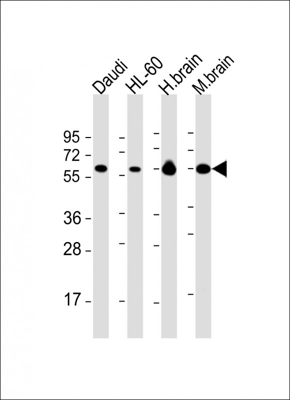
Figure 1. Western blot analysis of PACSIN2 using anti-PACSIN2 antibody (A04211-2). Electrophoresis was performed on a 5-20% SDS-PAGE gel at 70V (Stacking gel) / 90V (Resolving gel) for 2-3 hours. The sample well of each lane was loaded with 30 ug of sample under reducing conditions. Lane 1: human T-47D whole cell lysates, Lane 2: monkey COS-7 whole cell lysates, Lane 3: human placenta tissue lysates, Lane 4: human HepG2 whole cell lysates, Lane 5: rat lung tissue lysates, Lane 6: rat heart tissue tissue lysates, Lane 7: mouse lung tissue lysates, Lane 8: mouse heart tissue lysates. After electrophoresis, proteins were transferred to a nitrocellulose membrane at 150 mA for 50-90 minutes. Blocked the membrane with 5% non-fat milk/TBS for 1.5 hour at RT. The membrane was incubated with rabbit anti-PACSIN2 antigen affinity purified polyclonal antibody (Catalog # A04211-2) at 0.25 microg/mL overnight at 4°C, then washed with TBS-0.1%Tween 3 times with 5 minutes each and probed with a goat anti-rabbit IgG-HRP secondary antibody at a dilution of 1:5000 for 1.5 hour at RT. The signal is developed using an Enhanced Chemiluminescent detection (ECL) kit (Catalog # EK1002) with Tanon 5200 system. A specific band was detected for PACSIN2 at approximately 60 kDa. The expected band size for PACSIN2 is at 56,60-65 kDa.
. PACSIN2 was detected in a paraffin-embedded section of Diffuse large B-cell lymphoma of human intestine tissue. Heat mediated antigen retrieval was performed in EDTA buffer (pH 8.0, epitope retrieval solution). The tissue section was blocked with 10% goat serum. The tissue section was then incubated with 2 microg/ml rabbit anti-PACSIN2 Antibody (A04211-2) overnight at 4°C. Peroxidase Conjugated Goat Anti-rabbit IgG was used as secondary antibody and incubated for 30 minutes at 37°C. The tissue section was developed using HRP Conjugated Rabbit IgG Super Vision Assay Kit (Catalog # SV0002) with DAB as the chromogen.")
. PACSIN2 was detected in a paraffin-embedded section of human colon adenocarcinoma tissue. Heat mediated antigen retrieval was performed in EDTA buffer (pH 8.0, epitope retrieval solution). The tissue section was blocked with 10% goat serum. The tissue section was then incubated with 2 microg/ml rabbit anti-PACSIN2 Antibody (A04211-2) overnight at 4°C. Peroxidase Conjugated Goat Anti-rabbit IgG was used as secondary antibody and incubated for 30 minutes at 37°C. The tissue section was developed using HRP Conjugated Rabbit IgG Super Vision Assay Kit (Catalog # SV0002) with DAB as the chromogen.")
. PACSIN2 was detected in a paraffin-embedded section of human placenta tissue. Heat mediated antigen retrieval was performed in EDTA buffer (pH 8.0, epitope retrieval solution). The tissue section was blocked with 10% goat serum. The tissue section was then incubated with 2 microg/ml rabbit anti-PACSIN2 Antibody (A04211-2) overnight at 4°C. Peroxidase Conjugated Goat Anti-rabbit IgG was used as secondary antibody and incubated for 30 minutes at 37°C. The tissue section was developed using HRP Conjugated Rabbit IgG Super Vision Assay Kit (Catalog # SV0002) with DAB as the chromogen.")
. PACSIN2 was detected in a paraffin-embedded section of human testicular seminoma tissue. Heat mediated antigen retrieval was performed in EDTA buffer (pH 8.0, epitope retrieval solution). The tissue section was blocked with 10% goat serum. The tissue section was then incubated with 2 microg/ml rabbit anti-PACSIN2 Antibody (A04211-2) overnight at 4°C. Peroxidase Conjugated Goat Anti-rabbit IgG was used as secondary antibody and incubated for 30 minutes at 37°C. The tissue section was developed using HRP Conjugated Rabbit IgG Super Vision Assay Kit (Catalog # SV0002) with DAB as the chromogen.")
. PACSIN2 was detected in an immunocytochemical section of U2OS cells. Enzyme antigen retrieval was performed using IHC enzyme antigen retrieval reagent (AR0022) for 15 mins. The cells were blocked with 10% goat serum. And then incubated with 5 microg/mL rabbit anti-PACSIN2 Antibody (A04211-2) overnight at 4°C. Cy3 Conjugated Goat Anti-Rabbit IgG (BA1032) was used as secondary antibody at 1:500 dilution and incubated for 30 minutes at 37°C. The section was counterstained with DAPI. Visualize using a fluorescence microscope and filter sets appropriate for the label used.")
. PACSIN2 was detected in a paraffin-embedded section of human lung cancer tissue. Heat mediated antigen retrieval was performed in EDTA buffer (pH 8.0, epitope retrieval solution). The tissue section was blocked with 10% goat serum. The tissue section was then incubated with 5 microg/mL rabbit anti-PACSIN2 Antibody (A04211-2) overnight at 4°C. DyLight®550 Conjugated Goat Anti-Rabbit IgG (BA1135) was used as secondary antibody at 1:500 dilution and incubated for 30 minutes at 37°C. The section was counterstained with DAPI. Visualize using a fluorescence microscope and filter sets appropriate for the label used.")
. PACSIN2 was detected in a paraffin-embedded section of human testicular cancer tissue. Heat mediated antigen retrieval was performed in EDTA buffer (pH 8.0, epitope retrieval solution). The tissue section was blocked with 10% goat serum. The tissue section was then incubated with 5 microg/mL rabbit anti-PACSIN2 Antibody (A04211-2) overnight at 4°C. DyLight®550 Conjugated Goat Anti-Rabbit IgG (BA1135) was used as secondary antibody at 1:500 dilution and incubated for 30 minutes at 37°C. The section was counterstained with DAPI. Visualize using a fluorescence microscope and filter sets appropriate for the label used.")
. Overlay histogram showing Hela cells stained with A04211-2 (Blue line). To facilitate intracellular staining, cells were fixed with 4% paraformaldehyde and permeabilized with permeabilization buffer. The cells were blocked with 10% normal goat serum. And then incubated with rabbit anti-PACSIN2 Antibody (A04211-2, 1 microg/1x106 cells) for 30 min at 20°C. DyLight®488 conjugated goat anti-rabbit IgG (BA1127, 5-10 microg/1x106 cells) was used as secondary antibody for 30 minutes at 20°C. Isotype control antibody (Green line) was rabbit IgG (1 microg/1x106) used under the same conditions. Unlabelled sample without incubation with primary antibody and secondary antibody (Red line) was used as a blank control.")
Figure 1. Western blot analysis of PACSIN2 using anti-PACSIN2 antibody (A04211-2). Electrophoresis was performed on a 5-20% SDS-PAGE gel at 70V (Stacking gel) / 90V (Resolving gel) for 2-3 hours. The sample well of each lane was loaded with 30 ug of sample under reducing conditions. Lane 1: human T-47D whole cell lysates, Lane 2: monkey COS-7 whole cell lysates, Lane 3: human placenta tissue lysates, Lane 4: human HepG2 whole cell lysates, Lane 5: rat lung tissue lysates, Lane 6: rat heart tissue tissue lysates, Lane 7: mouse lung tissue lysates, Lane 8: mouse heart tissue lysates. After electrophoresis, proteins were transferred to a nitrocellulose membrane at 150 mA for 50-90 minutes. Blocked the membrane with 5% non-fat milk/TBS for 1.5 hour at RT. The membrane was incubated with rabbit anti-PACSIN2 antigen affinity purified polyclonal antibody (Catalog # A04211-2) at 0.25 microg/mL overnight at 4°C, then washed with TBS-0.1%Tween 3 times with 5 minutes each and probed with a goat anti-rabbit IgG-HRP secondary antibody at a dilution of 1:5000 for 1.5 hour at RT. The signal is developed using an Enhanced Chemiluminescent detection (ECL) kit (Catalog # EK1002) with Tanon 5200 system. A specific band was detected for PACSIN2 at approximately 60 kDa. The expected band size for PACSIN2 is at 56,60-65 kDa.
Anti-PACSIN2 Antibody Picoband(r)
A04211-2-CARRIER-FREE
ApplicationsFlow Cytometry, ImmunoFluorescence, Western Blot, ELISA, ImmunoCytoChemistry, ImmunoHistoChemistry
Product group Antibodies
ReactivityHuman, Monkey, Mouse, Rat
TargetPACSIN2
Overview
- SupplierBoster Bio
- Product NameAnti-PACSIN2 Antibody Picoband(r)
- Delivery Days Customer9
- ApplicationsFlow Cytometry, ImmunoFluorescence, Western Blot, ELISA, ImmunoCytoChemistry, ImmunoHistoChemistry
- CertificationResearch Use Only
- ClonalityPolyclonal
- Concentration500 ug/ml
- Gene ID11252
- Target namePACSIN2
- Target descriptionprotein kinase C and casein kinase substrate in neurons 2
- Target synonymsSDPII, protein kinase C and casein kinase substrate in neurons protein 2, cytoplasmic phosphoprotein PACSIN2, syndapin-2, syndapin-II, syndapin2
- HostRabbit
- IsotypeIgG
- Protein IDQ9UNF0
- Protein NameProtein kinase C and casein kinase substrate in neurons protein 2
- Scientific DescriptionBoster Bio Anti-PACSIN2 Antibody Picoband® catalog # A04211-2. Tested in ELISA, Flow Cytometry, IF, IHC, ICC, WB applications. This antibody reacts with Human, Mouse, Rat, Monkey. The brand Picoband indicates this is a premium antibody that guarantees superior quality, high affinity, and strong signals with minimal background in Western blot applications. Only our best-performing antibodies are designated as Picoband, ensuring unmatched performance.
- ReactivityHuman, Monkey, Mouse, Rat
- Storage Instruction-20°C,2°C to 8°C
- UNSPSC12352203
Related products
Product group Antibodies
Anti-PACSIN2 Antibody102-20258
ApplicationsWestern Blot
TargetPACSIN2
- SizePrice
Product group Antibodies
PACSIN2 Polyclonal AntibodyCAC15670
ApplicationsImmunoFluorescence, ImmunoPrecipitation, Western Blot, ELISA, ImmunoHistoChemistry
TargetPACSIN2
- SizePrice
Product group Antibodies
PACSIN2 Monoclonal AntibodyBSM-51639M
ApplicationsWestern Blot
ReactivityHuman, Mouse
TargetPACSIN2
- SizePrice
Product group Antibodies
[KO Validated] PACSIN2 AntibodyCAB19941
ReactivityHuman
TargetPACSIN2
- SizePrice

Product group Antibodies
Anti-PACSIN2 AntibodyHPA049854
ApplicationsImmunoCytoChemistry, ImmunoHistoChemistry
ReactivityHuman
TargetPACSIN2
- SizePrice
Product group Antibodies
PACSIN2 AntibodyCSB-PA793216
ApplicationsELISA, ImmunoHistoChemistry
ReactivityHuman, Mouse, Rat
TargetPACSIN2
- SizePrice
Product group Antibodies

PACSIN2 AntibodyLS-C409330
ApplicationsWestern Blot, ImmunoHistoChemistry
ReactivityHuman, Mouse
TargetPACSIN2
- SizePrice
![PACSIN2 antibody [N2C3] detects PACSIN2 protein at cytosol on human colon carcinoma by immunohistochemical analysis. Sample: Paraffin-embedded human colon carcinoma. PACSIN2 antibody [N2C3] (GTX104204) dilution: 1:500.
Antigen Retrieval: Trilogy? (EDTA based, pH 8.0) buffer, 15min](https://www.genetex.com/upload/website/prouct_img/normal/GTX104204/GTX104204_39918_IHC_w_23060120_185.webp)
Product group Antibodies
References
PACSIN2 antibody [N2C3]GTX104204
ApplicationsWestern Blot, ImmunoHistoChemistry, ImmunoHistoChemistry Paraffin
ReactivityHuman
TargetPACSIN2
- SizePrice
Product group Antibodies
Anti-PACSIN2 AntibodyA308485
ApplicationsWestern Blot
ReactivityHuman
- SizePrice