

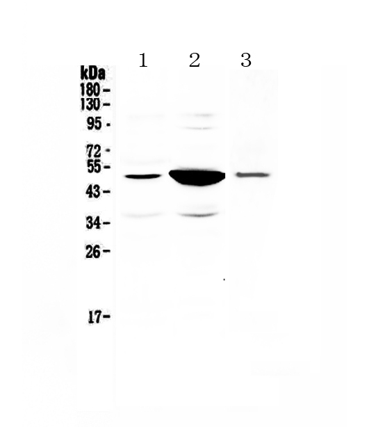
Figure 1. Western blot analysis of PAI1 using anti-PAI1 antibody (A00637-2). Electrophoresis was performed on a 5-20% SDS-PAGE gel at 70V (Stacking gel) / 90V (Resolving gel) for 2-3 hours. The sample well of each lane was loaded with 50ug of sample under reducing conditions. Lane 1: rat small intestine tissue lysates, Lane 2: rat kidney tissue lysates, Lane 3: mouse kidney tissue lysates. After Electrophoresis, proteins were transferred to a Nitrocellulose membrane at 150mA for 50-90 minutes. Blocked the membrane with 5% Non-fat Milk/ TBS for 1.5 hour at RT. The membrane was incubated with rabbit anti-PAI1 antigen affinity purified polyclonal antibody (Catalog # A00637-2) at 0.5 microg/mL overnight at 4°C, then washed with TBS-0.1%Tween 3 times with 5 minutes each and probed with a goat anti-rabbit IgG-HRP secondary antibody at a dilution of 1:10000 for 1.5 hour at RT. The signal is developed using an Enhanced Chemiluminescent detection (ECL) kit (Catalog # EK1002) with Tanon 5200 system. A specific band was detected for PAI1 at approximately 45-50KD. The expected band size for PAI1 is at 45KD.
Figure 1. Western blot analysis of PAI1 using anti-PAI1 antibody (A00637-2). Electrophoresis was performed on a 5-20% SDS-PAGE gel at 70V (Stacking gel) / 90V (Resolving gel) for 2-3 hours. The sample well of each lane was loaded with 50ug of sample under reducing conditions. Lane 1: rat small intestine tissue lysates, Lane 2: rat kidney tissue lysates, Lane 3: mouse kidney tissue lysates. After Electrophoresis, proteins were transferred to a Nitrocellulose membrane at 150mA for 50-90 minutes. Blocked the membrane with 5% Non-fat Milk/ TBS for 1.5 hour at RT. The membrane was incubated with rabbit anti-PAI1 antigen affinity purified polyclonal antibody (Catalog # A00637-2) at 0.5 microg/mL overnight at 4°C, then washed with TBS-0.1%Tween 3 times with 5 minutes each and probed with a goat anti-rabbit IgG-HRP secondary antibody at a dilution of 1:10000 for 1.5 hour at RT. The signal is developed using an Enhanced Chemiluminescent detection (ECL) kit (Catalog # EK1002) with Tanon 5200 system. A specific band was detected for PAI1 at approximately 45-50KD. The expected band size for PAI1 is at 45KD.
Anti-PAI1/Serpine1 Antibody Picoband(r)
A00637-2-CARRIER-FREE
ApplicationsWestern Blot, ELISA
Product group Antibodies
ReactivityMouse, Rat
TargetSERPINE1
Overview
- SupplierBoster Bio
- Product NameAnti-PAI1/Serpine1 Antibody Picoband(r)
- Delivery Days Customer9
- ApplicationsWestern Blot, ELISA
- CertificationResearch Use Only
- ClonalityPolyclonal
- Concentration500 ug/ml
- Gene ID5054
- Target nameSERPINE1
- Target descriptionserpin family E member 1
- Target synonymsPAI, PAI-1, PAI1, PLANH1, plasminogen activator inhibitor 1, endothelial plasminogen activator inhibitor, serine (or cysteine) proteinase inhibitor, clade E (nexin, plasminogen activator inhibitor type 1), member 1, serpin E1, serpin peptidase inhibitor, clade E (nexin, plasminogen activator inhibitor type 1), member 1
- HostRabbit
- IsotypeIgG
- Protein IDP20961
- Protein NamePlasminogen activator inhibitor 1
- Scientific DescriptionBoster Bio Anti-PAI1/Serpine1 Antibody Picoband® catalog # A00637-2. Tested in ELISA, WB applications. This antibody reacts with Mouse, Rat. The brand Picoband indicates this is a premium antibody that guarantees superior quality, high affinity, and strong signals with minimal background in Western blot applications. Only our best-performing antibodies are designated as Picoband, ensuring unmatched performance.
- ReactivityMouse, Rat
- Storage Instruction-20°C,2°C to 8°C
- UNSPSC12352203
Related products
Product group Antibodies
Anti-PAI1 AntibodyA15108
ApplicationsImmunoFluorescence, Western Blot, ImmunoCytoChemistry
ReactivityHuman, Mouse, Rat
- SizePrice
Product group Antibodies

Anti-PAI-1 [8H9D4]Ab03641-1.1
ApplicationsELISA, Neutralisation/Blocking
ReactivityHuman, Porcine
TargetSERPINE1
- SizePrice
Product group Antibodies
PAI1 Polyclonal AntibodyBS-6562R
ApplicationsImmunoFluorescence, ELISA, ImmunoCytoChemistry, ImmunoHistoChemistry, ImmunoHistoChemistry Frozen, ImmunoHistoChemistry Paraffin
ReactivityBovine, Canine, Equine, Guinea Pig, Human, Mouse, Porcine, Rat, Sheep
TargetSERPINE1
- SizePrice
Product group Antibodies
SERPINE1 AntibodyCSB-PA003699
ApplicationsWestern Blot, ELISA, ImmunoHistoChemistry
ReactivityHuman, Mouse, Rat
TargetSERPINE1
- SizePrice
Product group Antibodies

ApplicationsWestern Blot, ELISA, ImmunoHistoChemistry
ReactivityHuman
TargetSERPINE1
- SizePrice
Product group Antibodies
Serpine1 Polyclonal AntibodyCAC09137
ApplicationsImmunoFluorescence, Western Blot, ELISA, ImmunoHistoChemistry
TargetSERPINE1
- SizePrice
Product group Antibodies

ApplicationsELISA
ReactivityHuman
TargetSERPINE1
- SizePrice
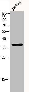
![WB analysis of HepG2 whole cell lysate using GTX02817 PAI-1 antibody [GT1223]. Dilution : 1:1000 Loading : 25μg](https://www.genetex.com/upload/website/prouct_img/normal/GTX02817/CutImage_A19096_WB_01_(1069081)_w_23053122_189.webp)
Product group Antibodies
PAI-1 antibody [GT1223]GTX02817
ApplicationsWestern Blot
ReactivityHuman
TargetSERPINE1
- SizePrice

Product group Antibodies
Anti-SERPINE1 AntibodyHPA050039
ApplicationsImmunoCytoChemistry, ImmunoHistoChemistry
ReactivityHuman
TargetSERPINE1
- SizePrice