


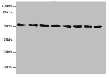
Figure 1. Western blot analysis of PDIA6 using anti-PDIA6 antibody (A03813-2). Electrophoresis was performed on a 5-20% SDS-PAGE gel at 70V (Stacking gel) / 90V (Resolving gel) for 2-3 hours. The sample well of each lane was loaded with 30ug of sample under reducing conditions. Lane 1: human K562 whole cell lysates, Lane 2: human HepG2 whole cell lysates, Lane 3: human HT1080 whole cell lysates, Lane 4: human Raji whole cell lysates, Lane 5: human Hela whole cell lysates, Lane 6: human HEK293 whole cell lysates, Lane 7: human THP-1 whole cell lysates, Lane 8: human SH-SY5Y whole cell lysates, Lane 9: rat kidney tissue lysates, Lane 10: rat heart tissue lysates, Lane 11: rat liver tissue lysates, Lane 12: rat lung tissue lysates, Lane 13: mouse kidney tissue lysates, Lane 14: mouse heart tissue lysates, Lane 15: mouse liver tissue lysates, Lane 16: mouse lung tissue lysates. After Electrophoresis, proteins were transferred to a Nitrocellulose membrane at 150mA for 50-90 minutes. Blocked the membrane with 5% Non-fat Milk/ TBS for 1.5 hour at RT. The membrane was incubated with rabbit anti-PDIA6 antigen affinity purified polyclonal antibody (Catalog # A03813-2) at 0.25 microg/mL overnight at 4°C, then washed with TBS-0.1%Tween 3 times with 5 minutes each and probed with a goat anti-rabbit IgG-HRP secondary antibody at a dilution of 1:5000 for 1.5 hour at RT. The signal is developed using an Enhanced Chemiluminescent detection (ECL) kit (Catalog # EK1002) with Tanon 5200 system. A specific band was detected for PDIA6 at approximately 50KD. The expected band size for PDIA6 is at 50KD.
. PDIA6 was detected in paraffin-embedded section of mouse ovary tissue. Heat mediated antigen retrieval was performed in EDTA buffer (pH8.0, epitope retrieval solution). The tissue section was blocked with 10% goat serum. The tissue section was then incubated with 2microg/ml rabbit anti-PDIA6 Antibody (A03813-2) overnight at 4°C. Biotinylated goat anti-rabbit IgG was used as secondary antibody and incubated for 30 minutes at 37°C. The tissue section was developed using Strepavidin-Biotin-Complex (SABC) (Catalog # SA1022) with DAB as the chromogen.")
. PDIA6 was detected in paraffin-embedded section of rat ovary tissue. Heat mediated antigen retrieval was performed in EDTA buffer (pH8.0, epitope retrieval solution). The tissue section was blocked with 10% goat serum. The tissue section was then incubated with 2microg/ml rabbit anti-PDIA6 Antibody (A03813-2) overnight at 4°C. Biotinylated goat anti-rabbit IgG was used as secondary antibody and incubated for 30 minutes at 37°C. The tissue section was developed using Strepavidin-Biotin-Complex (SABC) (Catalog # SA1022) with DAB as the chromogen.")
. PDIA6 was detected in paraffin-embedded section of human breast cancer tissue. Heat mediated antigen retrieval was performed in EDTA buffer (pH8.0, epitope retrieval solution). The tissue section was blocked with 10% goat serum. The tissue section was then incubated with 2microg/ml rabbit anti-PDIA6 Antibody (A03813-2) overnight at 4°C. Biotinylated goat anti-rabbit IgG was used as secondary antibody and incubated for 30 minutes at 37°C. The tissue section was developed using Strepavidin-Biotin-Complex (SABC) (Catalog # SA1022) with DAB as the chromogen.")
. PDIA6 was detected in paraffin-embedded section of human liver cancer tissue. Heat mediated antigen retrieval was performed in EDTA buffer (pH8.0, epitope retrieval solution). The tissue section was blocked with 10% goat serum. The tissue section was then incubated with 2microg/ml rabbit anti-PDIA6 Antibody (A03813-2) overnight at 4°C. Biotinylated goat anti-rabbit IgG was used as secondary antibody and incubated for 30 minutes at 37°C. The tissue section was developed using Strepavidin-Biotin-Complex (SABC) (Catalog # SA1022) with DAB as the chromogen.")
. PDIA6 was detected in paraffin-embedded section of human rectal cancer tissue. Heat mediated antigen retrieval was performed in EDTA buffer (pH8.0, epitope retrieval solution). The tissue section was blocked with 10% goat serum. The tissue section was then incubated with 2microg/ml rabbit anti-PDIA6 Antibody (A03813-2) overnight at 4°C. Biotinylated goat anti-rabbit IgG was used as secondary antibody and incubated for 30 minutes at 37°C. The tissue section was developed using Strepavidin-Biotin-Complex (SABC) (Catalog # SA1022) with DAB as the chromogen.")
. PDIA6 was detected in immunocytochemical section of U20S cells. Enzyme antigen retrieval was performed using IHC enzyme antigen retrieval reagent (AR0022) for 15 mins. The cells were blocked with 10% goat serum. And then incubated with 5microg/mL rabbit anti-PDIA6 Antibody (A03813-2) overnight at 4°C. DyLight®488 Conjugated Goat Anti-Rabbit IgG (BA1127) was used as secondary antibody at 1:100 dilution and incubated for 30 minutes at 37°C. The section was counterstained with DAPI. Visualize using a fluorescence microscope and filter sets appropriate for the label used.")
. Overlay histogram showing K562 cells stained with A03813-2 (Blue line). To facilitate intracellular staining, cells were fixed with 4% paraformaldehyde and permeabilized with permeabilization buffer. The cells were blocked with 10% normal goat serum. And then incubated with rabbit anti-PDIA6 Antibody (A03813-2, 1microg/1x106 cells) for 30 min at 20°C. DyLight®488 conjugated goat anti-rabbit IgG (BA1127, 5-10microg/1x106 cells) was used as secondary antibody for 30 minutes at 20°C. Isotype control antibody (Green line) was rabbit IgG (1microg/1x106) used under the same conditions. Unlabelled sample without incubation with primary antibody and secondary antibody (Red line) was used as a blank control.")
Figure 1. Western blot analysis of PDIA6 using anti-PDIA6 antibody (A03813-2). Electrophoresis was performed on a 5-20% SDS-PAGE gel at 70V (Stacking gel) / 90V (Resolving gel) for 2-3 hours. The sample well of each lane was loaded with 30ug of sample under reducing conditions. Lane 1: human K562 whole cell lysates, Lane 2: human HepG2 whole cell lysates, Lane 3: human HT1080 whole cell lysates, Lane 4: human Raji whole cell lysates, Lane 5: human Hela whole cell lysates, Lane 6: human HEK293 whole cell lysates, Lane 7: human THP-1 whole cell lysates, Lane 8: human SH-SY5Y whole cell lysates, Lane 9: rat kidney tissue lysates, Lane 10: rat heart tissue lysates, Lane 11: rat liver tissue lysates, Lane 12: rat lung tissue lysates, Lane 13: mouse kidney tissue lysates, Lane 14: mouse heart tissue lysates, Lane 15: mouse liver tissue lysates, Lane 16: mouse lung tissue lysates. After Electrophoresis, proteins were transferred to a Nitrocellulose membrane at 150mA for 50-90 minutes. Blocked the membrane with 5% Non-fat Milk/ TBS for 1.5 hour at RT. The membrane was incubated with rabbit anti-PDIA6 antigen affinity purified polyclonal antibody (Catalog # A03813-2) at 0.25 microg/mL overnight at 4°C, then washed with TBS-0.1%Tween 3 times with 5 minutes each and probed with a goat anti-rabbit IgG-HRP secondary antibody at a dilution of 1:5000 for 1.5 hour at RT. The signal is developed using an Enhanced Chemiluminescent detection (ECL) kit (Catalog # EK1002) with Tanon 5200 system. A specific band was detected for PDIA6 at approximately 50KD. The expected band size for PDIA6 is at 50KD.
Anti-PDIA6 Antibody Picoband(r)
A03813-2-CARRIER-FREE
ApplicationsFlow Cytometry, ImmunoFluorescence, Western Blot, ELISA, ImmunoCytoChemistry, ImmunoHistoChemistry
Product group Antibodies
ReactivityHuman, Mouse, Rat
TargetPDIA6
Overview
- SupplierBoster Bio
- Product NameAnti-PDIA6 Antibody Picoband(r)
- Delivery Days Customer9
- ApplicationsFlow Cytometry, ImmunoFluorescence, Western Blot, ELISA, ImmunoCytoChemistry, ImmunoHistoChemistry
- CertificationResearch Use Only
- ClonalityPolyclonal
- Concentration500 ug/ml
- Gene ID10130
- Target namePDIA6
- Target descriptionprotein disulfide isomerase family A member 6
- Target synonymsERP5, P5, TXNDC7, protein disulfide-isomerase A6, ER protein 5, endoplasmic reticulum protein 5, epididymis secretory sperm binding protein, protein disulfide isomerase P5, protein disulfide isomerase-associated 6, protein disulfide isomerase-related protein, thioredoxin domain containing 7 (protein disulfide isomerase), thioredoxin domain-containing protein 7
- HostRabbit
- IsotypeIgG
- Protein IDQ15084
- Protein NameProtein disulfide-isomerase A6
- Scientific DescriptionBoster Bio Anti-PDIA6 Antibody Picoband® catalog # A03813-2. Tested in ELISA, Flow Cytometry, IF, IHC, ICC, WB applications. This antibody reacts with Human, Mouse, Rat. The brand Picoband indicates this is a premium antibody that guarantees superior quality, high affinity, and strong signals with minimal background in Western blot applications. Only our best-performing antibodies are designated as Picoband, ensuring unmatched performance.
- ReactivityHuman, Mouse, Rat
- Storage Instruction-20°C,2°C to 8°C
- UNSPSC12352203
Related products
Product group Antibodies
Anti-PDIA6 AntibodyA31840
ApplicationsImmunoFluorescence, Western Blot, ImmunoHistoChemistry
ReactivityHuman, Mouse, Rat
- SizePrice
Product group Antibodies
Anti-PDIA6 Antibody144-07055
ApplicationsImmunoFluorescence, Western Blot, ImmunoHistoChemistry
ReactivityHuman, Mouse, Rat
TargetPDIA6
- SizePrice
Product group Antibodies
PDIA6 Recombinant Antibody, Biotin ConjugatedBSM-61392R-BIOTIN
ApplicationsWestern Blot, ImmunoHistoChemistry, ImmunoHistoChemistry Frozen, ImmunoHistoChemistry Paraffin
ReactivityHuman, Mouse, Rat
TargetPDIA6
- SizePrice
Product group Antibodies
PDIA6 AntibodyCSB-PA622988DSR1HU
ApplicationsWestern Blot, ELISA, ImmunoHistoChemistry
ReactivityHuman, Mouse
TargetPDIA6
- SizePrice
Product group Antibodies

PDIA6 / ERP5 AntibodyLS-C346205
ApplicationsImmunoFluorescence, Western Blot, ImmunoHistoChemistry
ReactivityHuman, Mouse, Rat
TargetPDIA6
- SizePrice
![PDIA6 antibody [N1N3] detects PDIA6 protein at cytoplasm in mouse liver by immunohistochemical analysis. Sample: Paraffin-embedded mouse liver. PDIA6 antibody [N1N3] (GTX121275) diluted at 1:500.
Antigen Retrieval: Citrate buffer, pH 6.0, 15 min](https://www.genetex.com/upload/website/prouct_img/normal/GTX121275/GTX121275_40464_20160803_IHC-P_M_w_23060519_402.webp)
Product group Antibodies
PDIA6 antibody [N1N3]GTX121275
ApplicationsImmunoPrecipitation, Western Blot, ImmunoHistoChemistry, ImmunoHistoChemistry Paraffin
ReactivityHuman, Mouse, Rat
TargetPDIA6
- SizePrice

Product group Antibodies
Anti-PDIA6 AntibodyHPA034652
ApplicationsWestern Blot, ImmunoHistoChemistry
ReactivityHuman
TargetPDIA6
- SizePrice