

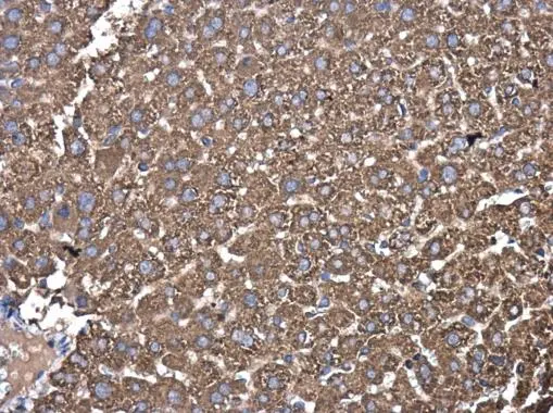
PDIA6 antibody [N1N3] detects PDIA6 protein at cytoplasm in mouse liver by immunohistochemical analysis. Sample: Paraffin-embedded mouse liver. PDIA6 antibody [N1N3] (GTX121275) diluted at 1:500.
Antigen Retrieval: Citrate buffer, pH 6.0, 15 min
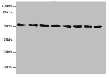
![Immunoprecipitation of PDIA6 protein from 293T whole cell extracts using 5 μg of PDIA6 antibody [N1N3] (GTX121275). Western blot analysis was performed using PDIA6 antibody [N1N3] (GTX121275). EasyBlot anti-Rabbit IgG (GTX221666-01) was used as a secondary reagent.](https://www.genetex.com/upload/website/prouct_img/normal/GTX121275/GTX121275_40464_20150420_IP_w_23060519_718.webp "Immunoprecipitation of PDIA6 protein from 293T whole cell extracts using 5 μg of PDIA6 antibody [N1N3] (GTX121275). Western blot analysis was performed using PDIA6 antibody [N1N3] (GTX121275). EasyBlot anti-Rabbit IgG (GTX221666-01) was used as a secondary reagent.")
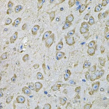
![PDIA6 antibody [N1N3] detects PDIA6 protein at cytoplasm in rat liver by immunohistochemical analysis. Sample: Paraffin-embedded rat liver. PDIA6 antibody [N1N3] (GTX121275) diluted at 1:500.
Antigen Retrieval: Citrate buffer, pH 6.0, 15 min](https://www.genetex.com/upload/website/prouct_img/normal/GTX121275/GTX121275_40464_20160803_IHC-P_R_w_23060519_914.webp "PDIA6 antibody [N1N3] detects PDIA6 protein at cytoplasm in rat liver by immunohistochemical analysis. Sample: Paraffin-embedded rat liver. PDIA6 antibody [N1N3] (GTX121275) diluted at 1:500.
Antigen Retrieval: Citrate buffer, pH 6.0, 15 min")
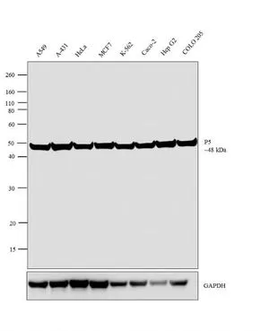
 A: Raji 10% SDS PAGE GTX121275 diluted at 1:1000")
PDIA6 antibody [N1N3] detects PDIA6 protein at cytoplasm in mouse liver by immunohistochemical analysis. Sample: Paraffin-embedded mouse liver. PDIA6 antibody [N1N3] (GTX121275) diluted at 1:500.
Antigen Retrieval: Citrate buffer, pH 6.0, 15 min
PDIA6 antibody [N1N3]
GTX121275
ApplicationsImmunoPrecipitation, Western Blot, ImmunoHistoChemistry, ImmunoHistoChemistry Paraffin
Product group Antibodies
ReactivityHuman, Mouse, Rat
TargetPDIA6
Overview
- SupplierGeneTex
- Product NamePDIA6 antibody [N1N3]
- Delivery Days Customer9
- Application Supplier NoteWB: 1:500-1:3000. IHC-P: 1:100-1:1000. IP: 1:100-1:500. *Optimal dilutions/concentrations should be determined by the researcher.Not tested in other applications.
- ApplicationsImmunoPrecipitation, Western Blot, ImmunoHistoChemistry, ImmunoHistoChemistry Paraffin
- CertificationResearch Use Only
- ClonalityPolyclonal
- Concentration1 mg/ml
- ConjugateUnconjugated
- Gene ID10130
- Target namePDIA6
- Target descriptionprotein disulfide isomerase family A member 6
- Target synonymsERP5, P5, TXNDC7, protein disulfide-isomerase A6, ER protein 5, endoplasmic reticulum protein 5, epididymis secretory sperm binding protein, protein disulfide isomerase P5, protein disulfide isomerase-associated 6, protein disulfide isomerase-related protein, thioredoxin domain containing 7 (protein disulfide isomerase), thioredoxin domain-containing protein 7
- HostRabbit
- IsotypeIgG
- Protein IDQ15084
- Protein NameProtein disulfide-isomerase A6
- Scientific DescriptionProtein disulfide isomerases (EC 5.3.4.1), such as PDIA6, are endoplasmic reticulum (ER) resident proteins that catalyze formation, reduction, and isomerization of disulfide bonds in proteins and are thought to play a role in folding of disulfide-bonded proteins (Hayano and Kikuchi, 1995 [PubMed 7590364]).[supplied by OMIM]
- ReactivityHuman, Mouse, Rat
- Storage Instruction-20°C or -80°C,2°C to 8°C
- UNSPSC41116161
Datasheet
Related products
Product group Antibodies
Anti-PDIA6 AntibodyA31840
ApplicationsImmunoFluorescence, Western Blot, ImmunoHistoChemistry
ReactivityHuman, Mouse, Rat
- SizePrice

Product group Antibodies
Anti-PDIA6 Antibody Picoband(r)A03813-2-CARRIER-FREE
ApplicationsFlow Cytometry, ImmunoFluorescence, Western Blot, ELISA, ImmunoCytoChemistry, ImmunoHistoChemistry
ReactivityHuman, Mouse, Rat
TargetPDIA6
- SizePrice
Product group Antibodies
Anti-PDIA6 Antibody144-07055
ApplicationsImmunoFluorescence, Western Blot, ImmunoHistoChemistry
ReactivityHuman, Mouse, Rat
TargetPDIA6
- SizePrice
Product group Antibodies
PDIA6 Recombinant Antibody, Biotin ConjugatedBSM-61392R-BIOTIN
ApplicationsWestern Blot, ImmunoHistoChemistry, ImmunoHistoChemistry Frozen, ImmunoHistoChemistry Paraffin
ReactivityHuman, Mouse, Rat
TargetPDIA6
- SizePrice
Product group Antibodies
PDIA6 AntibodyCSB-PA622988DSR1HU
ApplicationsWestern Blot, ELISA, ImmunoHistoChemistry
ReactivityHuman, Mouse
TargetPDIA6
- SizePrice
Product group Antibodies

PDIA6 / ERP5 AntibodyLS-C346205
ApplicationsImmunoFluorescence, Western Blot, ImmunoHistoChemistry
ReactivityHuman, Mouse, Rat
TargetPDIA6
- SizePrice
Product group Antibodies
PDIA6 antibodyGTX11432
ApplicationsFlow Cytometry, ImmunoFluorescence, Western Blot, ImmunoCytoChemistry
ReactivityHuman, Porcine
TargetPDIA6
- SizePrice
Product group Antibodies
PDIA6 antibodyGTX33397
ApplicationsImmunoFluorescence, Western Blot, ImmunoCytoChemistry, ImmunoHistoChemistry, ImmunoHistoChemistry Paraffin
ReactivityHuman, Mouse, Rat
TargetPDIA6
- SizePrice

Product group Antibodies
Anti-PDIA6 AntibodyHPA034652
ApplicationsWestern Blot, ImmunoHistoChemistry
ReactivityHuman
TargetPDIA6
- SizePrice