

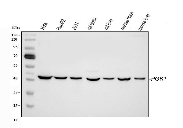
Figure 1. Western blot analysis of PGK1 using anti-PGK1 antibody (PB9774). Electrophoresis was performed on a 5-20% SDS-PAGE gel at 70V (Stacking gel) / 90V (Resolving gel) for 2-3 hours. The sample well of each lane was loaded with 30 ug of sample under reducing conditions. Lane 1: human Hela whole cell lysates, Lane 2: human HepG2 whole cell lysates, Lane 3: human 293T whole cell lysates, Lane 4: rat brain tissue lysates, Lane 5: rat liver tissue lysates, Lane 6: mouse brain tissue lysates, Lane 7: mouse liver tissue lysates. After electrophoresis, proteins were transferred to a nitrocellulose membrane at 150 mA for 50-90 minutes. Blocked the membrane with 5% non-fat milk/TBS for 1.5 hour at RT. The membrane was incubated with rabbit anti-PGK1 antigen affinity purified polyclonal antibody (Catalog # PB9774) at 0.5 microg/mL overnight at 4°C, then washed with TBS-0.1%Tween 3 times with 5 minutes each and probed with a goat anti-rabbit IgG-HRP secondary antibody at a dilution of 1:5000 for 1.5 hour at RT. The signal is developed using an Enhanced Chemiluminescent detection (ECL) kit (Catalog # EK1002) with Tanon 5200 system. A specific band was detected for PGK1 at approximately 43 kDa. The expected band size for PGK1 is at 43 kDa.
. PGK1 was detected in an immunocytochemical section of U87 cells. Enzyme antigen retrieval was performed using IHC enzyme antigen retrieval reagent (AR0022) for 15 mins. The cells were blocked with 10% goat serum. And then incubated with 5 microg/mL rabbit anti-PGK1 Antibody (PB9774) overnight at 4°C. Cy3 Conjugated Goat Anti-Rabbit IgG (BA1032) was used as secondary antibody at 1:500 dilution and incubated for 30 minutes at 37°C. Visualize using a fluorescence microscope and filter sets appropriate for the label used.")
. Overlay histogram showing 293T cells stained with PB9774 (Blue line). To facilitate intracellular staining, cells were fixed with 4% paraformaldehyde and permeabilized with permeabilization buffer. The cells were blocked with 10% normal goat serum. And then incubated with rabbit anti-PGK1 Antibody (PB9774, 1 microg/1x106 cells) for 30 min at 20°C. DyLight®488 conjugated goat anti-rabbit IgG (BA1127, 5-10 microg/1x106 cells) was used as secondary antibody for 30 minutes at 20°C. Isotype control antibody (Green line) was rabbit IgG (1 microg/1x106) used under the same conditions. Unlabelled sample without incubation with primary antibody and secondary antibody (Red line) was used as a blank control.")
Figure 1. Western blot analysis of PGK1 using anti-PGK1 antibody (PB9774). Electrophoresis was performed on a 5-20% SDS-PAGE gel at 70V (Stacking gel) / 90V (Resolving gel) for 2-3 hours. The sample well of each lane was loaded with 30 ug of sample under reducing conditions. Lane 1: human Hela whole cell lysates, Lane 2: human HepG2 whole cell lysates, Lane 3: human 293T whole cell lysates, Lane 4: rat brain tissue lysates, Lane 5: rat liver tissue lysates, Lane 6: mouse brain tissue lysates, Lane 7: mouse liver tissue lysates. After electrophoresis, proteins were transferred to a nitrocellulose membrane at 150 mA for 50-90 minutes. Blocked the membrane with 5% non-fat milk/TBS for 1.5 hour at RT. The membrane was incubated with rabbit anti-PGK1 antigen affinity purified polyclonal antibody (Catalog # PB9774) at 0.5 microg/mL overnight at 4°C, then washed with TBS-0.1%Tween 3 times with 5 minutes each and probed with a goat anti-rabbit IgG-HRP secondary antibody at a dilution of 1:5000 for 1.5 hour at RT. The signal is developed using an Enhanced Chemiluminescent detection (ECL) kit (Catalog # EK1002) with Tanon 5200 system. A specific band was detected for PGK1 at approximately 43 kDa. The expected band size for PGK1 is at 43 kDa.
Anti-PGK1 Antibody Picoband(r)
PB9774-CARRIER-FREE
ApplicationsFlow Cytometry, ImmunoFluorescence, Western Blot, ImmunoCytoChemistry
Product group Antibodies
ReactivityHuman, Mouse, Rat
TargetPGK1
Overview
- SupplierBoster Bio
- Product NameAnti-PGK1 Antibody Picoband(r)
- Delivery Days Customer9
- Application Supplier NoteTested Species: In-house tested species with positive results. Other applications have not been tested. Optimal dilutions should be determined by end users.
- ApplicationsFlow Cytometry, ImmunoFluorescence, Western Blot, ImmunoCytoChemistry
- CertificationResearch Use Only
- ClonalityPolyclonal
- Concentration500 ug/ml
- Gene ID5230
- Target namePGK1
- Target descriptionphosphoglycerate kinase 1
- Target synonymsHEL-S-68p, MIG10, PGKA, phosphoglycerate kinase 1, PRP 2, cell migration-inducing gene 10 protein, epididymis secretory sperm binding protein Li 68p, primer recognition protein 2
- HostRabbit
- IsotypeIgG
- Protein IDP00558
- Protein NamePhosphoglycerate kinase 1
- Scientific DescriptionBoster Bio Anti-PGK1 Antibody Picoband® catalog # PB9774. Tested in Flow Cytometry, IF, ICC, WB applications. This antibody reacts with Human, Mouse, Rat. The brand Picoband indicates this is a premium antibody that guarantees superior quality, high affinity, and strong signals with minimal background in Western blot applications. Only our best-performing antibodies are designated as Picoband, ensuring unmatched performance.
- ReactivityHuman, Mouse, Rat
- Storage Instruction-20°C,2°C to 8°C
- UNSPSC12352203
Related products
Product group Antibodies
Anti-PGK1 AntibodyA29944
ApplicationsImmunoFluorescence, Western Blot, ImmunoHistoChemistry
ReactivityHuman, Mouse, Rat
- SizePrice
Product group Antibodies
Anti-PGK1 Antibody144-60560
ApplicationsWestern Blot, ImmunoHistoChemistry
ReactivityHuman, Mouse, Rat
TargetPGK1
- SizePrice
Product group Antibodies
PGK1 Recombinant AntibodyBSM-61216R
ApplicationsFlow Cytometry, ImmunoFluorescence, Western Blot, ImmunoCytoChemistry
TargetPGK1
- SizePrice
Product group Antibodies

Goat anti-PGK1EB12390
ApplicationsWestern Blot, ELISA
ReactivityHuman
TargetPGK1
- SizePrice
Product group Antibodies
PGK1 AntibodyCSB-PA547426
ApplicationsWestern Blot, ELISA, ImmunoHistoChemistry
ReactivityHuman, Mouse, Rat
TargetPGK1
- SizePrice
Product group Antibodies
Pgk1 Polyclonal AntibodyCAC07097
ApplicationsWestern Blot, ELISA
TargetPGK1
- SizePrice
Product group Antibodies

PGK1 / Phosphoglycerate Kinase AntibodyLS-C401396
ApplicationsWestern Blot, ELISA, ImmunoHistoChemistry
ReactivityHuman, Mouse, Rat
TargetPGK1
- SizePrice
Product group Antibodies
PGK1 antibody [N1C1]GTX101405
ApplicationsWestern Blot, ImmunoHistoChemistry, ImmunoHistoChemistry Paraffin
ReactivityHuman, Mouse, Rat
TargetPGK1
- SizePrice

Product group Antibodies
Anti-PGK1 AntibodyHPA045385
ApplicationsImmunoHistoChemistry
ReactivityHuman
TargetPGK1
- SizePrice