


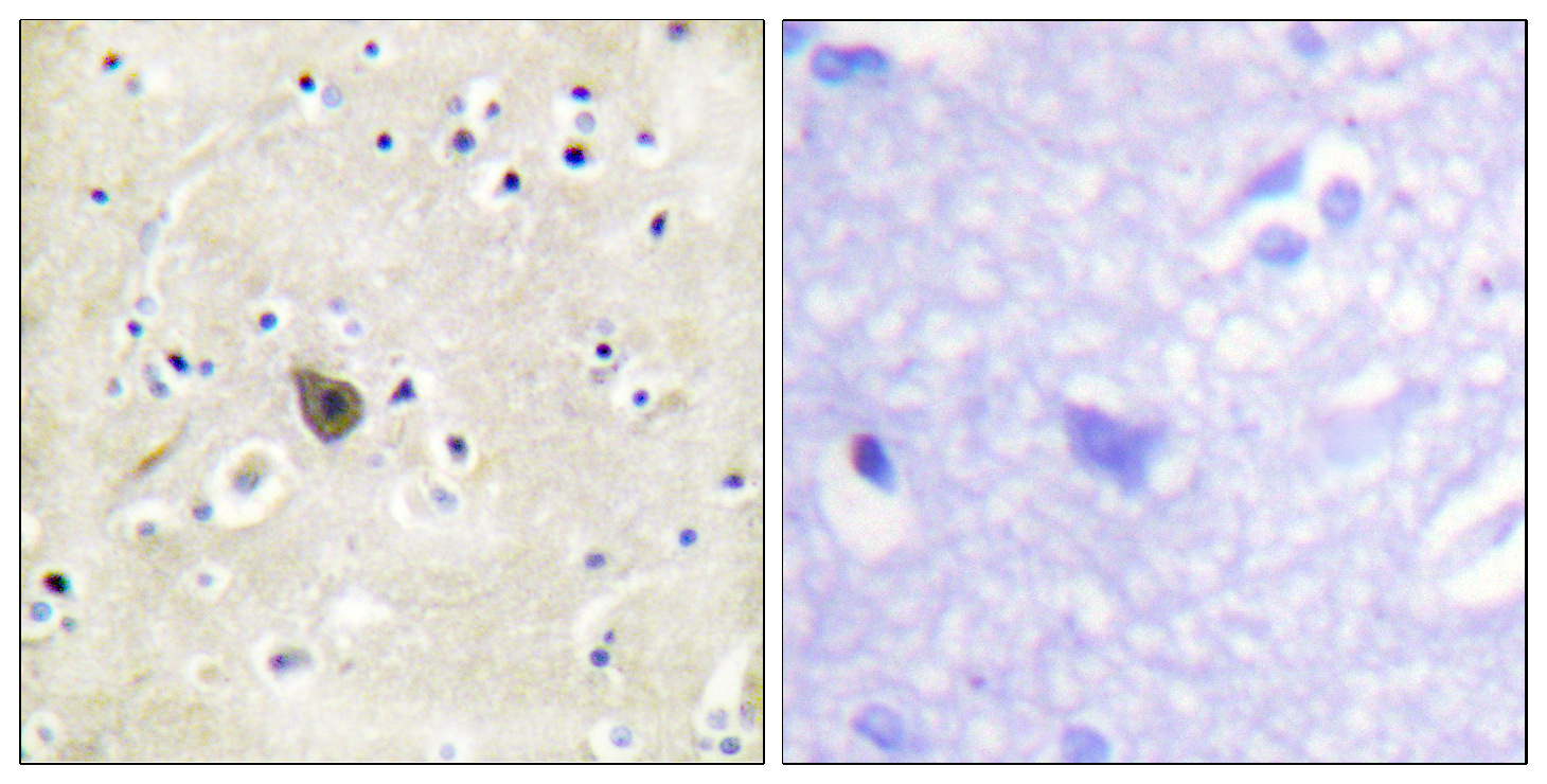
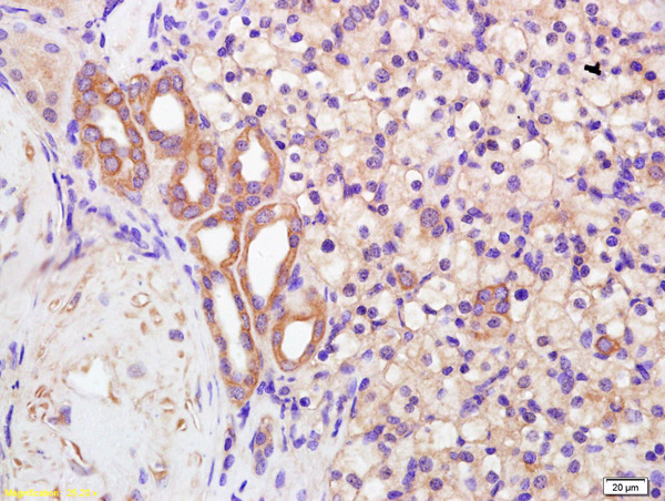
Immunohistochemical staining of human kidney shows cytoplasmic positivity in cells in tubules.

Immunohistochemical staining of human kidney shows cytoplasmic positivity in cells in tubules.
Anti-PKD2 Antibody
HPA015794
ApplicationsImmunoCytoChemistry, ImmunoHistoChemistry
Product group Antibodies
ReactivityHuman
TargetPKD2
Overview
- SupplierAtlas Antibodies
- Product NameAnti-PKD2 Antibody
- Delivery Days Customer4
- ApplicationsImmunoCytoChemistry, ImmunoHistoChemistry
- CertificationResearch Use Only
- ClonalityPolyclonal
- ConjugateUnconjugated
- Gene ID5311
- Target namePKD2
- Target descriptionpolycystin 2, transient receptor potential cation channel
- Target synonymsAPKD2, PC2, PKD4, Pc-2, TRPP2, polycystin-2, autosomal dominant polycystic kidney disease type II protein, polycystic kidney disease 2 (autosomal dominant), transient receptor potential cation channel subfamily P member 2
- HostRabbit
- IsotypeIgG
- Protein IDQ13563
- Protein NamePolycystin-2
- Scientific DescriptionRecombinant Protein Epitope Signature Tag (PrEST) antigen sequence
- ReactivityHuman
- Storage Instruction-20°C,2°C to 8°C
- UNSPSC41116161
Datasheet
MSDS
Related products
Product group Antibodies
Anti-PKD2 AntibodyA96142
ApplicationsWestern Blot, ELISA, ImmunoHistoChemistry
ReactivityHuman, Mouse, Rat
- SizePrice

Product group Antibodies
Anti-Polycystin 2/PKD2 Antibody Picoband(r)A00630-3-CARRIER-FREE
ApplicationsFlow Cytometry, Western Blot, ELISA, ImmunoHistoChemistry
ReactivityHuman, Mouse, Rat
TargetPKD2
- SizePrice
Product group Antibodies
Anti-PKD2 Antibody144-65722
ApplicationsWestern Blot
ReactivityHuman, Mouse, Rat
TargetPKD2
- SizePrice
Product group Antibodies
Polycystin 2 Polyclonal AntibodyBS-2158R
ApplicationsImmunoFluorescence, ELISA, ImmunoCytoChemistry, ImmunoHistoChemistry, ImmunoHistoChemistry Frozen, ImmunoHistoChemistry Paraffin
ReactivityBovine, Canine, Chicken, Human, Mouse, Rat
TargetPKD2
- SizePrice
Product group Antibodies

ApplicationsWestern Blot, ELISA
ReactivityBovine, Human, Mouse, Rat
TargetPKD2
- SizePrice
Product group Antibodies
PKD2 Polyclonal AntibodyCAC12932
ApplicationsImmunoFluorescence, ELISA, ImmunoHistoChemistry
TargetPKD2
- SizePrice
Product group Antibodies
Phospho-PKD2 (S812) AntibodyCSB-PA030064
ApplicationsImmunoFluorescence, ELISA
ReactivityHuman, Mouse, Rat
TargetPKD2
- SizePrice
Product group Antibodies

PKD2 / Polycystin 2 AntibodyLS-C332683
ApplicationsWestern Blot
ReactivityHuman, Mouse, Rat
TargetPKD2
- SizePrice
Product group Antibodies
Polycystin 2 antibodyGTX113802
ApplicationsWestern Blot, ImmunoHistoChemistry, ImmunoHistoChemistry Paraffin
ReactivityHuman, Zebra Fish
TargetPKD2
- SizePrice