


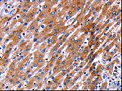
Immunohistochemical staining of human testis shows strong nuclear positivity in cells in seminiferous ducts.
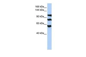
![Lane 1: Marker [kDa] 230, 130, 95, 72, 56, 36, 28, 17, 11. Lane 2: Human cell line RT-4. Lane 3: Human cell line U-251MG sp](https://atlasantibodies.s3.amazonaws.com/images/wb/hpa006394-wb-1.jpg "Lane 1: Marker [kDa] 230, 130, 95, 72, 56, 36, 28, 17, 11. Lane 2: Human cell line RT-4. Lane 3: Human cell line U-251MG sp")
Immunohistochemical staining of human testis shows strong nuclear positivity in cells in seminiferous ducts.
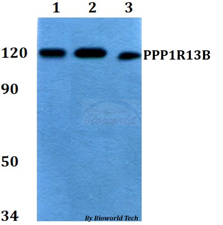
Anti-PPP1R13B Antibody
HPA006394
ApplicationsWestern Blot, ImmunoCytoChemistry, ImmunoHistoChemistry
Product group Antibodies
ReactivityHuman, Mouse, Rat
TargetPPP1R13B
Overview
- SupplierAtlas Antibodies
- Product NameAnti-PPP1R13B Antibody
- Delivery Days Customer4
- ApplicationsWestern Blot, ImmunoCytoChemistry, ImmunoHistoChemistry
- CertificationResearch Use Only
- ClonalityPolyclonal
- ConjugateUnconjugated
- Gene ID23368
- Target namePPP1R13B
- Target descriptionprotein phosphatase 1 regulatory subunit 13B
- Target synonymsASPP1, p53BP2-like, p85, apoptosis-stimulating of p53 protein 1, apoptosis-stimulating protein of p53, 1, protein phosphatase 1, regulatory (inhibitor) subunit 13B
- HostRabbit
- IsotypeIgG
- Protein IDQ96KQ4
- Protein NameApoptosis-stimulating of p53 protein 1
- Scientific DescriptionRecombinant Protein Epitope Signature Tag (PrEST) antigen sequence
- ReactivityHuman, Mouse, Rat
- Storage Instruction-20°C,2°C to 8°C
- UNSPSC41116161
Datasheet
MSDS
Related products
Product group Antibodies
PPP1R13B AntibodyCSB-PA071472
ApplicationsELISA, ImmunoHistoChemistry
ReactivityHuman, Mouse
TargetPPP1R13B
- SizePrice

Product group Antibodies
Anti-PPP1R13B Antibody Picoband(r)A07257-2-CARRIER-FREE
ApplicationsWestern Blot, ELISA
ReactivityHuman, Mouse, Rat
TargetPPP1R13B
- SizePrice
Product group Antibodies
Anti-PPP1R13B AntibodyA28927
ApplicationsWestern Blot
ReactivityHuman, Mouse, Rat
- SizePrice
Product group Antibodies

PPP1R13B AntibodyLS-C750380
ApplicationsWestern Blot
ReactivityHuman, Mouse, Rat
TargetPPP1R13B
- SizePrice
Product group Antibodies
ASPP1 antibody, InternalGTX46274
ApplicationsWestern Blot
ReactivityHuman
TargetPPP1R13B
- SizePrice
Product group Antibodies
Anti-PPP1R13B (Center) Antibody102-22630
ApplicationsWestern Blot
TargetPPP1R13B
- SizePrice
Product group Antibodies
References
ASPP1 Polyclonal AntibodyBS-1282R
ApplicationsImmunoFluorescence, Western Blot, ELISA, ImmunoCytoChemistry, ImmunoHistoChemistry, ImmunoHistoChemistry Frozen, ImmunoHistoChemistry Paraffin
ReactivityBovine, Canine, Chicken, Equine, Human, Mouse, Rat
TargetPPP1R13B
- SizePrice