

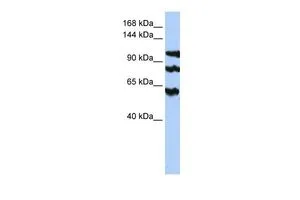
WB analysis of HepG2 cells using GTX46274 PPP1R13B antibody at 0.2-1μg/ml.
WB analysis of HepG2 cells using GTX46274 PPP1R13B antibody at 0.2-1μg/ml.
ASPP1 antibody, Internal
GTX46274
ApplicationsWestern Blot
Product group Antibodies
ReactivityHuman
TargetPPP1R13B
Overview
- SupplierGeneTex
- Product NameASPP1 antibody, Internal
- Delivery Days Customer9
- Application Supplier NoteWB: 0.2-2.5 ug/ml. *Optimal dilutions/concentrations should be determined by the researcher.Not tested in other applications.
- ApplicationsWestern Blot
- CertificationResearch Use Only
- ClonalityPolyclonal
- Concentration0.5-1 mg/ml
- ConjugateUnconjugated
- Gene ID23368
- Target namePPP1R13B
- Target descriptionprotein phosphatase 1 regulatory subunit 13B
- Target synonymsASPP1, p53BP2-like, p85, apoptosis-stimulating of p53 protein 1, apoptosis-stimulating protein of p53, 1, protein phosphatase 1, regulatory (inhibitor) subunit 13B
- HostRabbit
- IsotypeIgG
- Protein IDQ96KQ4
- Protein NameApoptosis-stimulating of p53 protein 1
- Scientific DescriptionThis gene encodes a member of the ASPP (apoptosis-stimulating protein of p53) family of p53 interacting proteins. The protein contains four ankyrin repeats and an SH3 domain involved in protein-protein interactions. ASPP proteins are required for the induction of apoptosis by p53-family proteins. They promote DNA binding and transactivation of p53-family proteins on the promoters of proapoptotic genes. Expression of this gene is regulated by the E2F transcription factor. [provided by RefSeq, Jul 2008]
- ReactivityHuman
- Storage Instruction-20°C or -80°C,2°C to 8°C
- UNSPSC41116161
Datasheet
Related products
Product group Antibodies
PPP1R13B AntibodyCSB-PA071472
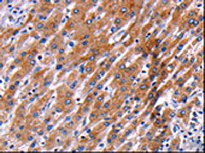
ApplicationsELISA, ImmunoHistoChemistry
ReactivityHuman, Mouse
TargetPPP1R13B
- SizePrice

Product group Antibodies
Anti-PPP1R13B Antibody Picoband(r)A07257-2-CARRIER-FREE
ApplicationsWestern Blot, ELISA
ReactivityHuman, Mouse, Rat
TargetPPP1R13B
- SizePrice
Product group Antibodies
Anti-PPP1R13B AntibodyA28927
ApplicationsWestern Blot
ReactivityHuman, Mouse, Rat
- SizePrice
Product group Antibodies

PPP1R13B AntibodyLS-C750380
ApplicationsWestern Blot
ReactivityHuman, Mouse, Rat
TargetPPP1R13B
- SizePrice

Product group Antibodies
Anti-PPP1R13B AntibodyHPA006394
ApplicationsWestern Blot, ImmunoCytoChemistry, ImmunoHistoChemistry
ReactivityHuman, Mouse, Rat
TargetPPP1R13B
- SizePrice
Product group Antibodies
ASPP1 antibodyGTX48719
ApplicationsWestern Blot, ELISA
ReactivityHuman
TargetPPP1R13B
- SizePrice
Product group Antibodies
Anti-PPP1R13B (Center) Antibody102-22630
ApplicationsWestern Blot
TargetPPP1R13B
- SizePrice
Product group Antibodies
References
ASPP1 Polyclonal AntibodyBS-1282R
ApplicationsImmunoFluorescence, Western Blot, ELISA, ImmunoCytoChemistry, ImmunoHistoChemistry, ImmunoHistoChemistry Frozen, ImmunoHistoChemistry Paraffin
ReactivityBovine, Canine, Chicken, Equine, Human, Mouse, Rat
TargetPPP1R13B
- SizePrice