


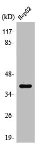
Figure 1. Western blot analysis of PU.1/SPI1 using anti-PU.1/SPI1 antibody (A01116-1). Electrophoresis was performed on a 5-20% SDS-PAGE gel at 70V (Stacking gel) / 90V (Resolving gel) for 2-3 hours. The sample well of each lane was loaded with 30 ug of sample under reducing conditions. Lane 1: human K562 whole cell lysates, Lane 2: human Raji whole cell lysates. After electrophoresis, proteins were transferred to a nitrocellulose membrane at 150 mA for 50-90 minutes. Blocked the membrane with 5% non-fat milk/TBS for 1.5 hour at RT. The membrane was incubated with rabbit anti-PU.1/SPI1 antigen affinity purified polyclonal antibody (Catalog # A01116-1) at 0.5 microg/mL overnight at 4°C, then washed with TBS-0.1%Tween 3 times with 5 minutes each and probed with a goat anti-rabbit IgG-HRP secondary antibody at a dilution of 1:5000 for 1.5 hour at RT. The signal is developed using an Enhanced Chemiluminescent detection (ECL) kit (Catalog # EK1002) with Tanon 5200 system. A specific band was detected for PU.1/SPI1 at approximately 40 kDa. The expected band size for PU.1/SPI1 is at 40 kDa.
. PU.1/SPI1 was detected in a paraffin-embedded section of human lymphoma tissue. Heat mediated antigen retrieval was performed in EDTA buffer (pH 8.0, epitope retrieval solution). The tissue section was blocked with 10% goat serum. The tissue section was then incubated with 2 microg/ml rabbit anti-PU.1/SPI1 Antibody (A01116-1) overnight at 4°C. Peroxidase Conjugated Goat Anti-rabbit IgG was used as secondary antibody and incubated for 30 minutes at 37°C. The tissue section was developed using HRP Conjugated Rabbit IgG Super Vision Assay Kit (Catalog # SV0002) with DAB as the chromogen.")
. PU.1/SPI1 was detected in a paraffin-embedded section of mouse lymph gland tissue. Heat mediated antigen retrieval was performed in EDTA buffer (pH 8.0, epitope retrieval solution). The tissue section was blocked with 10% goat serum. The tissue section was then incubated with 2 microg/ml rabbit anti-PU.1/SPI1 Antibody (A01116-1) overnight at 4°C. Peroxidase Conjugated Goat Anti-rabbit IgG was used as secondary antibody and incubated for 30 minutes at 37°C. The tissue section was developed using HRP Conjugated Rabbit IgG Super Vision Assay Kit (Catalog # SV0002) with DAB as the chromogen.")
. PU.1/SPI1 was detected in a paraffin-embedded section of rat lymph gland tissue. Heat mediated antigen retrieval was performed in EDTA buffer (pH 8.0, epitope retrieval solution). The tissue section was blocked with 10% goat serum. The tissue section was then incubated with 2 microg/ml rabbit anti-PU.1/SPI1 Antibody (A01116-1) overnight at 4°C. Peroxidase Conjugated Goat Anti-rabbit IgG was used as secondary antibody and incubated for 30 minutes at 37°C. The tissue section was developed using HRP Conjugated Rabbit IgG Super Vision Assay Kit (Catalog # SV0002) with DAB as the chromogen.")
. Overlay histogram showing THP-1 cells stained with A01116-1 (Blue line). To facilitate intracellular staining, cells were fixed with 4% paraformaldehyde and permeabilized with permeabilization buffer. The cells were blocked with 10% normal goat serum. And then incubated with rabbit anti-PU.1/SPI1 Antibody (A01116-1, 1 microg/1x106 cells) for 30 min at 20°C. DyLight®488 conjugated goat anti-rabbit IgG (BA1127, 5-10 microg/1x106 cells) was used as secondary antibody for 30 minutes at 20°C. Isotype control antibody (Green line) was rabbit IgG (1 microg/1x106) used under the same conditions. Unlabelled sample without incubation with primary antibody and secondary antibody (Red line) was used as a blank control.")
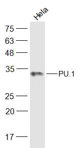
Figure 1. Western blot analysis of PU.1/SPI1 using anti-PU.1/SPI1 antibody (A01116-1). Electrophoresis was performed on a 5-20% SDS-PAGE gel at 70V (Stacking gel) / 90V (Resolving gel) for 2-3 hours. The sample well of each lane was loaded with 30 ug of sample under reducing conditions. Lane 1: human K562 whole cell lysates, Lane 2: human Raji whole cell lysates. After electrophoresis, proteins were transferred to a nitrocellulose membrane at 150 mA for 50-90 minutes. Blocked the membrane with 5% non-fat milk/TBS for 1.5 hour at RT. The membrane was incubated with rabbit anti-PU.1/SPI1 antigen affinity purified polyclonal antibody (Catalog # A01116-1) at 0.5 microg/mL overnight at 4°C, then washed with TBS-0.1%Tween 3 times with 5 minutes each and probed with a goat anti-rabbit IgG-HRP secondary antibody at a dilution of 1:5000 for 1.5 hour at RT. The signal is developed using an Enhanced Chemiluminescent detection (ECL) kit (Catalog # EK1002) with Tanon 5200 system. A specific band was detected for PU.1/SPI1 at approximately 40 kDa. The expected band size for PU.1/SPI1 is at 40 kDa.
Anti-PU.1/SPI1 Antibody Picoband(r)
A01116-1-CARRIER-FREE
ApplicationsFlow Cytometry, Western Blot, ELISA, ImmunoHistoChemistry
Product group Antibodies
ReactivityHuman, Mouse, Rat
TargetSPI1
Overview
- SupplierBoster Bio
- Product NameAnti-PU.1/SPI1 Antibody Picoband(r)
- Delivery Days Customer9
- ApplicationsFlow Cytometry, Western Blot, ELISA, ImmunoHistoChemistry
- CertificationResearch Use Only
- ClonalityPolyclonal
- Concentration500 ug/ml
- Gene ID6688
- Target nameSPI1
- Target descriptionSpi-1 proto-oncogene
- Target synonymsAGM10, OF, PU.1, SFPI1, SPI-1, SPI-A, transcription factor PU.1, 31 kDa transforming protein, hematopoietic transcription factor PU.1, spleen focus forming virus (SFFV) proviral integration oncogene spi1
- HostRabbit
- IsotypeIgG
- Protein IDP17947
- Protein NameTranscription factor PU.1
- Scientific DescriptionBoster Bio Anti-PU.1/SPI1 Antibody Picoband® catalog # A01116-1. Tested in ELISA, Flow Cytometry, IHC, WB applications. This antibody reacts with Human, Mouse, Rat. The brand Picoband indicates this is a premium antibody that guarantees superior quality, high affinity, and strong signals with minimal background in Western blot applications. Only our best-performing antibodies are designated as Picoband, ensuring unmatched performance.
- ReactivityHuman, Mouse, Rat
- Storage Instruction-20°C,2°C to 8°C
- UNSPSC12352203
Related products
Product group Antibodies
SPI1 AntibodyCSB-PA003887
ApplicationsImmunoFluorescence, Western Blot, ELISA, ImmunoHistoChemistry
ReactivityHuman, Monkey, Mouse, Rat
TargetSPI1
- SizePrice

Product group Antibodies
Anti-SPI1 AntibodyA97901
ApplicationsWestern Blot, ELISA
ReactivityHuman, Mouse, Rat
- SizePrice
Product group Antibodies

SPI1 / PU.1 AntibodyLS-C831700
ApplicationsWestern Blot, ELISA
ReactivityHuman, Mouse, Rat
TargetSPI1
- SizePrice
Product group Antibodies

Goat anti-PU.1EB08429
ApplicationsWestern Blot, ELISA, ImmunoHistoChemistry
ReactivityHuman, Rat
TargetSPI1
- SizePrice

Product group Antibodies
Anti-SPI1 AntibodyHPA044653
ApplicationsChIP Chromatin ImmunoPrecipitation, ImmunoCytoChemistry, ImmunoHistoChemistry
ReactivityHuman
TargetSPI1
- SizePrice
Product group Antibodies
PU.1/Spi1 Polyclonal AntibodyBS-19594R
ApplicationsImmunoFluorescence, Western Blot, ELISA, ImmunoCytoChemistry, ImmunoHistoChemistry, ImmunoHistoChemistry Frozen, ImmunoHistoChemistry Paraffin
ReactivityBovine, Equine, Human, Mouse, Porcine, Rat, Sheep
TargetSPI1
- SizePrice
![THP-1 whole cell and nuclear extracts (30 μg) were separated by 10% SDS-PAGE, and the membrane was blotted with PU.1 antibody [N1C1] (GTX101581) diluted at 1:500. The HRP-conjugated anti-rabbit IgG antibody (GTX213110-01) was used to detect the primary antibody.](https://www.genetex.com/upload/website/prouct_img/normal/GTX101581/GTX101581_43600_20190607_WB_Fraction_w_23060100_464.webp)
Product group Antibodies
PU.1 antibody [N1C1]GTX101581
ApplicationsWestern Blot
ReactivityHuman, Mouse
TargetSPI1
- SizePrice