


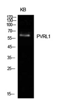
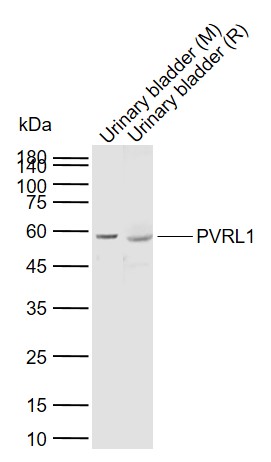
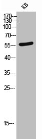
Figure 1. Western blot analysis of PVRL1/NECTIN1 using anti-PVRL1/NECTIN1 antibody (A04988-1). Electrophoresis was performed on a 5-20% SDS-PAGE gel at 70V (Stacking gel) / 90V (Resolving gel) for 2-3 hours. The sample well of each lane was loaded with 30ug of sample under reducing conditions. Lane 1: human Hek293 whole cell lysates, Lane 2: human CCRF-CEM whole cell lysates, Lane 3: human Sw620 whole cell lysates, Lane 4: human U87 whole cell lysates, Lane 5: human SGC-7901 whole cell lysates, Lane 6: human Jurkat whole cell lysates, Lane 7: rat PC-12 whole cell lysates, Lane 8: mouse spleen tissue lysates, Lane 9: mouse NIH/3T3 whole cell lysates, Lane 10: mouse Raw264.7 whole cell lysates. After Electrophoresis, proteins were transferred to a Nitrocellulose membrane at 150mA for 50-90 minutes. Blocked the membrane with 5% Non-fat Milk/ TBS for 1.5 hour at RT. The membrane was incubated with rabbit anti-PVRL1/NECTIN1 antigen affinity purified polyclonal antibody (Catalog # A04988-1) at 0.25 microg/mL overnight at 4°C, then washed with TBS-0.1%Tween 3 times with 5 minutes each and probed with a goat anti-rabbit IgG-HRP secondary antibody at a dilution of 1:5000 for 1.5 hour at RT. The signal is developed using an Enhanced Chemiluminescent detection (ECL) kit (Catalog # EK1002) with Tanon 5200 system. A specific band was detected for PVRL1/NECTIN1 at approximately 110KD. The expected band size for PVRL1/NECTIN1 is at 110KD.
. PVRL1/NECTIN1 was detected in paraffin-embedded section of human rectal cancer tissue. Heat mediated antigen retrieval was performed in EDTA buffer (pH8.0, epitope retrieval solution). The tissue section was blocked with 10% goat serum. The tissue section was then incubated with 1microg/ml rabbit anti-PVRL1/NECTIN1 Antibody (A04988-1) overnight at 4°C. Biotinylated goat anti-rabbit IgG was used as secondary antibody and incubated for 30 minutes at 37°C. The tissue section was developed using Strepavidin-Biotin-Complex (SABC) (Catalog # SA1022) with DAB as the chromogen.")
. PVRL1/NECTIN1 was detected in paraffin-embedded section of human rectal cancer tissue. Heat mediated antigen retrieval was performed in EDTA buffer (pH8.0, epitope retrieval solution). The tissue section was blocked with 10% goat serum. The tissue section was then incubated with 1microg/ml rabbit anti-PVRL1/NECTIN1 Antibody (A04988-1) overnight at 4°C. Biotinylated goat anti-rabbit IgG was used as secondary antibody and incubated for 30 minutes at 37°C. The tissue section was developed using Strepavidin-Biotin-Complex (SABC) (Catalog # SA1022) with DAB as the chromogen.")
. PVRL1/NECTIN1 was detected in paraffin-embedded section of human rectal cancer tissue. Heat mediated antigen retrieval was performed in EDTA buffer (pH8.0, epitope retrieval solution). The tissue section was blocked with 10% goat serum. The tissue section was then incubated with 1microg/ml rabbit anti-PVRL1/NECTIN1 Antibody (A04988-1) overnight at 4°C. Biotinylated goat anti-rabbit IgG was used as secondary antibody and incubated for 30 minutes at 37°C. The tissue section was developed using Strepavidin-Biotin-Complex (SABC) (Catalog # SA1022) with DAB as the chromogen.")
. PVRL1/NECTIN1 was detected in paraffin-embedded section of rat brain tissue. Heat mediated antigen retrieval was performed in EDTA buffer (pH8.0, epitope retrieval solution). The tissue section was blocked with 10% goat serum. The tissue section was then incubated with 1microg/ml rabbit anti-PVRL1/NECTIN1 Antibody (A04988-1) overnight at 4°C. Biotinylated goat anti-rabbit IgG was used as secondary antibody and incubated for 30 minutes at 37°C. The tissue section was developed using Strepavidin-Biotin-Complex (SABC) (Catalog # SA1022) with DAB as the chromogen.")
. PVRL1/NECTIN1 was detected in paraffin-embedded section of rat brain tissue. Heat mediated antigen retrieval was performed in EDTA buffer (pH8.0, epitope retrieval solution). The tissue section was blocked with 10% goat serum. The tissue section was then incubated with 1microg/ml rabbit anti-PVRL1/NECTIN1 Antibody (A04988-1) overnight at 4°C. Biotinylated goat anti-rabbit IgG was used as secondary antibody and incubated for 30 minutes at 37°C. The tissue section was developed using Strepavidin-Biotin-Complex (SABC) (Catalog # SA1022) with DAB as the chromogen.")
. Overlay histogram showing PC-3 cells stained with A04988-1 (Blue line). The cells were fixed with 4% paraformaldehyde and blocked with 10% normal goat serum. And then incubated with rabbit anti-PVRL1/NECTIN1 Antibody (A04988-1, 1microg/1x106 cells) for 30 min at 20°C. DyLight®488 conjugated goat anti-rabbit IgG (BA1127, 5-10microg/1x106 cells) was used as secondary antibody for 30 minutes at 20°C. Isotype control antibody (Green line) was rabbit IgG (1microg/1x106) used under the same conditions. Unlabelled sample without incubation with primary antibody and secondary antibody (Red line) was used as a blank control.")
Figure 1. Western blot analysis of PVRL1/NECTIN1 using anti-PVRL1/NECTIN1 antibody (A04988-1). Electrophoresis was performed on a 5-20% SDS-PAGE gel at 70V (Stacking gel) / 90V (Resolving gel) for 2-3 hours. The sample well of each lane was loaded with 30ug of sample under reducing conditions. Lane 1: human Hek293 whole cell lysates, Lane 2: human CCRF-CEM whole cell lysates, Lane 3: human Sw620 whole cell lysates, Lane 4: human U87 whole cell lysates, Lane 5: human SGC-7901 whole cell lysates, Lane 6: human Jurkat whole cell lysates, Lane 7: rat PC-12 whole cell lysates, Lane 8: mouse spleen tissue lysates, Lane 9: mouse NIH/3T3 whole cell lysates, Lane 10: mouse Raw264.7 whole cell lysates. After Electrophoresis, proteins were transferred to a Nitrocellulose membrane at 150mA for 50-90 minutes. Blocked the membrane with 5% Non-fat Milk/ TBS for 1.5 hour at RT. The membrane was incubated with rabbit anti-PVRL1/NECTIN1 antigen affinity purified polyclonal antibody (Catalog # A04988-1) at 0.25 microg/mL overnight at 4°C, then washed with TBS-0.1%Tween 3 times with 5 minutes each and probed with a goat anti-rabbit IgG-HRP secondary antibody at a dilution of 1:5000 for 1.5 hour at RT. The signal is developed using an Enhanced Chemiluminescent detection (ECL) kit (Catalog # EK1002) with Tanon 5200 system. A specific band was detected for PVRL1/NECTIN1 at approximately 110KD. The expected band size for PVRL1/NECTIN1 is at 110KD.
Anti-PVRL1/NECTIN1 Antibody Picoband(r)
A04988-1-CARRIER-FREE
ApplicationsFlow Cytometry, Western Blot, ELISA, ImmunoHistoChemistry
Product group Antibodies
ReactivityHuman, Mouse, Rat
TargetNECTIN1
Overview
- SupplierBoster Bio
- Product NameAnti-PVRL1/NECTIN1 Antibody Picoband(r)
- Delivery Days Customer9
- ApplicationsFlow Cytometry, Western Blot, ELISA, ImmunoHistoChemistry
- CertificationResearch Use Only
- ClonalityPolyclonal
- Concentration500 ug/ml
- Gene ID5818
- Target nameNECTIN1
- Target descriptionnectin cell adhesion molecule 1
- Target synonymsCD111, CLPED1, ED4, HIgR, HV1S, HVEC, OFC7, PRR, PRR1, PVRL1, PVRR, PVRR1, SK-12, nectin-1, nectin-1, ectodermal dysplasia 4 (Margarita Island type), herpes simplex virus type 1 sensitivity, herpes virus entry mediator C, herpesvirus Ig-like receptor, nectin 1, poliovirus receptor-like 1, poliovirus receptor-related 1 (herpesvirus entry mediator C), poliovirus receptor-related protein 1
- HostRabbit
- IsotypeIgG
- Protein IDQ15223
- Protein NameNectin-1
- Scientific DescriptionBoster Bio Anti-PVRL1/NECTIN1 Antibody Picoband® catalog # A04988-1. Tested in ELISA, Flow Cytometry, IHC, WB applications. This antibody reacts with Human, Mouse, Rat. The brand Picoband indicates this is a premium antibody that guarantees superior quality, high affinity, and strong signals with minimal background in Western blot applications. Only our best-performing antibodies are designated as Picoband, ensuring unmatched performance.
- ReactivityHuman, Mouse, Rat
- Storage Instruction-20°C,2°C to 8°C
- UNSPSC12352203
Related products
Product group Antibodies
Anti-PVRL1 AntibodyA97319
ApplicationsWestern Blot, ELISA
ReactivityHuman, Mouse, Rat
- SizePrice
Product group Antibodies
Anti-PVRL1 Antibody144-65455
ApplicationsWestern Blot
ReactivityHuman, Mouse, Rat
TargetNECTIN1
- SizePrice
Product group Antibodies
References
PVRL1 Polyclonal AntibodyBS-11126R
ApplicationsDot Blot, ImmunoFluorescence, Western Blot, ELISA, ImmunoCytoChemistry, ImmunoHistoChemistry, ImmunoHistoChemistry Frozen, ImmunoHistoChemistry Paraffin
TargetNECTIN1
- SizePrice
Product group Antibodies
PVRL1 AntibodyCSB-PA006247
ApplicationsWestern Blot, ELISA
ReactivityHuman, Mouse, Rat
TargetNECTIN1
- SizePrice
Product group Antibodies

Nectin-1 / PVRL1 AntibodyLS-C405545
ApplicationsWestern Blot, ELISA
ReactivityHuman, Mouse, Rat
TargetNECTIN1
- SizePrice

Product group Antibodies
Anti-NECTIN1 AntibodyHPA026846
ApplicationsImmunoHistoChemistry
ReactivityHuman
TargetNECTIN1
- SizePrice
![Nectin 1 antibody detects Nectin 1 protein by immunofluorescent analysis. Sample: DIV14 rat E18 primary cortical neurons were fixed in 4% paraformaldehyde at RT for 15 min. Green: Nectin 1 protein stained by Nectin 1 antibody (GTX130824) diluted at 1:500. Red: beta Tubulin 3/ Tuj1, stained by beta Tubulin 3/ Tuj1 antibody [GT1338] (GTX631831) diluted at 1:500. Blue: Fluoroshield with DAPI (GTX30920).](https://www.genetex.com/upload/website/prouct_img/normal/GTX130824/GTX130824_42144_20170719_IFA_R_w_23060523_921.webp)
Product group Antibodies
Nectin 1 antibodyGTX130824
ApplicationsImmunoFluorescence, Western Blot, ImmunoCytoChemistry
ReactivityHuman, Rat
TargetNECTIN1
- SizePrice
Product group Antibodies
Anti-HLA-DQB1 AntibodyCAB20372
ApplicationsWestern Blot, ELISA, ImmunoHistoChemistry, ImmunoHistoChemistry Paraffin
ReactivityHuman
TargetNECTIN1
- SizePrice