


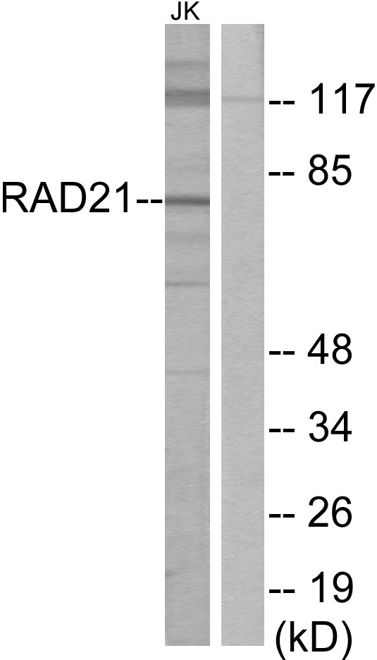
Figure 1. Western blot analysis of RAD21 using anti-RAD21 antibody (A01864-2). Electrophoresis was performed on a 5-20% SDS-PAGE gel at 70V (Stacking gel) / 90V (Resolving gel) for 2-3 hours. The sample well of each lane was loaded with 30 ug of sample under reducing conditions. Lane 1: monkey COS-7 whole cell lysates, Lane 2: human Hela whole cell lysates, Lane 3: human MOLT-4 whole cell lysates, Lane 4: human Jurkat whole cell lysates, Lane 5: human HEL whole cell lysates, Lane 6: human Daudi whole cell lysates, Lane 7: human A431 whole cell lysates, Lane 8: human T-47D whole cell lysates, Lane 9: rat brain tissue lysates, Lane 10: rat pancreas tissue lysates, Lane 11: mouse brain tissue lysates, Lane 12: mouse pancreas tissue lysates. After electrophoresis, proteins were transferred to a nitrocellulose membrane at 150 mA for 50-90 minutes. Blocked the membrane with 5% non-fat milk/TBS for 1.5 hour at RT. The membrane was incubated with rabbit anti-RAD21 antigen affinity purified polyclonal antibody (Catalog # A01864-2) at 0.5 microg/mL overnight at 4°C, then washed with TBS-0.1%Tween 3 times with 5 minutes each and probed with a goat anti-rabbit IgG-HRP secondary antibody at a dilution of 1:5000 for 1.5 hour at RT. The signal is developed using an Enhanced Chemiluminescent detection (ECL) kit (Catalog # EK1002) with Tanon 5200 system. A specific band was detected for RAD21 at approximately 72 kDa. The expected band size for RAD21 is at 72 kDa.
. RAD21 was detected in an immunocytochemical section of SiHa cells. Enzyme antigen retrieval was performed using IHC enzyme antigen retrieval reagent (AR0022) for 15 mins. The cells were blocked with 10% goat serum. And then incubated with 5 microg/mL rabbit anti-RAD21 Antibody (A01864-2) overnight at 4°C. DyLight®488 Conjugated Goat Anti-Rabbit IgG (BA1127) was used as secondary antibody at 1:100 dilution and incubated for 30 minutes at 37°C. The section was counterstained with DAPI. Visualize using a fluorescence microscope and filter sets appropriate for the label used.")
. Overlay histogram showing HL-60 cells stained with A01864-2 (Blue line). To facilitate intracellular staining, cells were fixed with 4% paraformaldehyde and permeabilized with permeabilization buffer. The cells were blocked with 10% normal goat serum. And then incubated with rabbit anti-RAD21 Antibody (A01864-2, 1 microg/1x106 cells) for 30 min at 20°C. DyLight®488 conjugated goat anti-rabbit IgG (BA1127, 5-10 microg/1x106 cells) was used as secondary antibody for 30 minutes at 20°C. Isotype control antibody (Green line) was rabbit IgG (1 microg/1x106) used under the same conditions. Unlabelled sample without incubation with primary antibody and secondary antibody (Red line) was used as a blank control.")
Figure 1. Western blot analysis of RAD21 using anti-RAD21 antibody (A01864-2). Electrophoresis was performed on a 5-20% SDS-PAGE gel at 70V (Stacking gel) / 90V (Resolving gel) for 2-3 hours. The sample well of each lane was loaded with 30 ug of sample under reducing conditions. Lane 1: monkey COS-7 whole cell lysates, Lane 2: human Hela whole cell lysates, Lane 3: human MOLT-4 whole cell lysates, Lane 4: human Jurkat whole cell lysates, Lane 5: human HEL whole cell lysates, Lane 6: human Daudi whole cell lysates, Lane 7: human A431 whole cell lysates, Lane 8: human T-47D whole cell lysates, Lane 9: rat brain tissue lysates, Lane 10: rat pancreas tissue lysates, Lane 11: mouse brain tissue lysates, Lane 12: mouse pancreas tissue lysates. After electrophoresis, proteins were transferred to a nitrocellulose membrane at 150 mA for 50-90 minutes. Blocked the membrane with 5% non-fat milk/TBS for 1.5 hour at RT. The membrane was incubated with rabbit anti-RAD21 antigen affinity purified polyclonal antibody (Catalog # A01864-2) at 0.5 microg/mL overnight at 4°C, then washed with TBS-0.1%Tween 3 times with 5 minutes each and probed with a goat anti-rabbit IgG-HRP secondary antibody at a dilution of 1:5000 for 1.5 hour at RT. The signal is developed using an Enhanced Chemiluminescent detection (ECL) kit (Catalog # EK1002) with Tanon 5200 system. A specific band was detected for RAD21 at approximately 72 kDa. The expected band size for RAD21 is at 72 kDa.
Anti-RAD21 Antibody Picoband(r)
A01864-2-DYLIGHT550
ApplicationsFlow Cytometry, ImmunoFluorescence, Western Blot, ELISA, ImmunoCytoChemistry
Product group Antibodies
ReactivityHuman, Monkey, Mouse, Rat
TargetRAD21
Overview
- SupplierBoster Bio
- Product NameAnti-RAD21 Antibody Picoband(r)
- Delivery Days Customer9
- ApplicationsFlow Cytometry, ImmunoFluorescence, Western Blot, ELISA, ImmunoCytoChemistry
- CertificationResearch Use Only
- ClonalityPolyclonal
- Concentration500 ug/ml
- ConjugateDyLight 550
- Gene ID5885
- Target nameRAD21
- Target descriptionRAD21 cohesin complex component
- Target synonymsCDLS4, HR21, HRAD21, MCD1, MGS, NXP1, SCC1, hHR21, double-strand-break repair protein rad21 homolog, NXP-1, RAD21 homolog, SCC1 homolog, kleisin, nuclear matrix protein 1, protein involved in DNA double-strand break repair, sister chromatid cohesion 1
- HostRabbit
- IsotypeIgG
- Protein IDO60216
- Protein NameDouble-strand-break repair protein rad21 homolog
- Scientific DescriptionBoster Bio Anti-RAD21 Antibody Picoband® catalog # A01864-2. Tested in ELISA, Flow Cytometry, IF, ICC, WB applications. This antibody reacts with Human, Mouse, Rat, Monkey. The brand Picoband indicates this is a premium antibody that guarantees superior quality, high affinity, and strong signals with minimal background in Western blot applications. Only our best-performing antibodies are designated as Picoband, ensuring unmatched performance.
- ReactivityHuman, Monkey, Mouse, Rat
- Storage Instruction-20°C,2°C to 8°C
- UNSPSC12352203
Related products
Product group Antibodies
References
Rad21 antibodyGTX106012
ApplicationsImmunoFluorescence, ImmunoPrecipitation, Western Blot, ChIP Chromatin ImmunoPrecipitation, ImmunoCytoChemistry, ImmunoHistoChemistry, ImmunoHistoChemistry Paraffin
ReactivityHuman, Mouse, Rat
TargetRAD21
- SizePrice
Product group Antibodies
Anti-RAD21 (C-term) Antibody102-25155
ApplicationsWestern Blot
TargetRAD21
- SizePrice
Product group Antibodies
Anti-RAD21 AntibodyA99001
ApplicationsWestern Blot, ELISA
ReactivityHuman, Mouse
- SizePrice
Product group Antibodies
Anti-RAD21 Antibody Picoband(r)A01864-2-CARRIER-FREE
ApplicationsFlow Cytometry, ImmunoFluorescence, Western Blot, ELISA, ImmunoCytoChemistry
ReactivityHuman, Monkey, Mouse, Rat
TargetRAD21
- SizePrice
Product group Antibodies
RAD21 AntibodyCSB-PA040139
ApplicationsImmunoFluorescence, Western Blot, ELISA, ImmunoHistoChemistry
ReactivityHuman, Mouse
TargetRAD21
- SizePrice
Product group Antibodies

RAD21 Antibody (C-Terminus)LS-C368680
ApplicationsImmunoFluorescence, Western Blot, ImmunoCytoChemistry, ImmunoHistoChemistry, ImmunoHistoChemistry Paraffin
ReactivityBovine, Human, Mouse, Rat, Zebra Fish
TargetRAD21
- SizePrice
Product group Antibodies
RAD21 Recombinant AntibodyBSM-61159R
ApplicationsFlow Cytometry, Western Blot
TargetRAD21
- SizePrice

Product group Antibodies
Anti-RAD21 AntibodyHPA020044
ApplicationsWestern Blot, ChIP Chromatin ImmunoPrecipitation, ImmunoCytoChemistry, ImmunoHistoChemistry
ReactivityHuman
TargetRAD21
- SizePrice