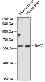
Bio-Connect
Anti-RAG2 Antibody

Anti-RAG2 Antibody

A90629
Antibodies.com
ApplicationsWestern Blot
Product group Antibodies
Sign in to order and to see your custom pricing.
Large volume orders?
Order with a bulk request

Overview

  • Supplier
    Antibodies.com
  • Product Name
    Anti-RAG2 Antibody
  • Delivery Days Customer
    7
  • Applications
    Western Blot
  • Certification
    Research Use Only
  • Clonality
    Polyclonal
  • Conjugate
    Unconjugated
  • Host
    Rabbit
  • Isotype
    IgG
  • Scientific Description
    Rabbit polyclonal antibody to RAG2.
  • UNSPSC
    12352203

Related products

Product group Antibodies
RayBiotech
ApplicationsWestern Blot
TargetRAG2
  • SizePrice
Product group Antibodies
Assay Genie
TargetRAG2
  • SizePrice
Product group Antibodies
Biosynth
RAG2 antibody70R-13300
  • SizePrice
Immunofluorescence staining of Hela cells with CSB-PA345128LA01HU at 1:100, counter-stained with DAPI. The cells were fixed in 4% formaldehyde, permeabilized using 0.2% Triton X-100 and blocked in 10% normal Goat Serum. The cells were then incubated with the antibody overnight at 4°C. The secondary antibody was Alexa Fluor 488-congugated AffiniPure Goat Anti-Rabbit IgG(H+L).
Product group Antibodies
Cusabio
RAG2 AntibodyCSB-PA345128LA01HU
ApplicationsImmunoFluorescence, ELISA
ReactivityHuman
TargetRAG2
  • SizePrice
Immunofluorescent staining of human cell line REH shows localization to nucleoplasm.
Product group Antibodies
Atlas Antibodies
ApplicationsImmunoCytoChemistry
ReactivityHuman
TargetRAG2
  • SizePrice
Figure 1. Western blot analysis of RAG2 using anti-RAG2 antibody (A00352). Electrophoresis was performed on a 5-20% SDS-PAGE gel at 70V (Stacking gel) / 90V (Resolving gel) for 2-3 hours. The sample well of each lane was loaded with 30 ug of sample under reducing conditions. Lane 1: human MOLT-4 whole cell lysates, Lane 2: mouse thymus tissue lysates, Lane 6: mouse spleen tissue lysates. After electrophoresis, proteins were transferred to a nitrocellulose membrane at 150 mA for 50-90 minutes. Blocked the membrane with 5% non-fat milk/TBS for 1.5 hour at RT. The membrane was incubated with rabbit anti-RAG2 antigen affinity purified polyclonal antibody (Catalog # A00352) at 0.5 microg/mL overnight at 4°C, then washed with TBS-0.1%Tween 3 times with 5 minutes each and probed with a goat anti-rabbit IgG-HRP secondary antibody at a dilution of 1:5000 for 1.5 hour at RT. The signal is developed using an Enhanced Chemiluminescent detection (ECL) kit (Catalog # EK1002) with Tanon 5200 system. A specific band was detected for RAG2 at approximately 55 kDa. The expected band size for RAG2 is at 59 kDa.
Product group Antibodies
Boster Bio
ApplicationsFlow Cytometry, Western Blot, ELISA
TargetRAG2
  • SizePrice
Product group Antibodies
GeneTex
RAG2 antibodyGTX109710
ApplicationsImmunoFluorescence, Western Blot, ImmunoCytoChemistry, ImmunoHistoChemistry, ImmunoHistoChemistry Paraffin
TargetRAG2
  • SizePrice
Product group Antibodies
Bioss Antibodies
ApplicationsImmunoFluorescence, ELISA, ImmunoCytoChemistry, ImmunoHistoChemistry, ImmunoHistoChemistry Frozen, ImmunoHistoChemistry Paraffin
TargetRAG2
  • SizePrice
Product group Antibodies
LifeSpan BioSciences
ApplicationsWestern Blot
TargetRAG2
  • SizePrice