

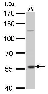
RAG2 antibody detects RAG2 protein by Western blot analysis. A. 50 μg Rat thymus lysate/extract 7.5 % SDS-PAGE RAG2 antibody (GTX109710) dilution: 1:1000
 A: Molt-4 (GTX27912) 7.5% SDS PAGE GTX109710 diluted at 1:1000")
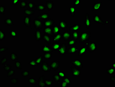
 diluted at 1:500. Red: Phalloidin, a cytoskeleton marker, diluted at 1:200. Scale bar = 10 μm.")
 antibody at 1:100 dilution.
Antigen Retrieval: Trilogy? (EDTA based, pH 8.0) buffer, 15min")
RAG2 antibody detects RAG2 protein by Western blot analysis. A. 50 μg Rat thymus lysate/extract 7.5 % SDS-PAGE RAG2 antibody (GTX109710) dilution: 1:1000
RAG2 antibody
GTX109710
ApplicationsImmunoFluorescence, Western Blot, ImmunoCytoChemistry, ImmunoHistoChemistry, ImmunoHistoChemistry Paraffin
Product group Antibodies
ReactivityHuman, Rat
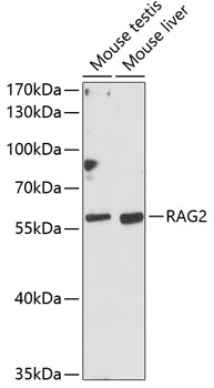
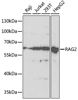
TargetRAG2
Overview
- SupplierGeneTex
- Product NameRAG2 antibody
- Delivery Days Customer9
- Application Supplier NoteWB: 1:500-1:3000. ICC/IF: 1:100-1:1000. IHC-P: 1:100-1:1000. *Optimal dilutions/concentrations should be determined by the researcher.Not tested in other applications.
- ApplicationsImmunoFluorescence, Western Blot, ImmunoCytoChemistry, ImmunoHistoChemistry, ImmunoHistoChemistry Paraffin
- CertificationResearch Use Only
- ClonalityPolyclonal
- Concentration1.01 mg/ml
- ConjugateUnconjugated
- Gene ID5897
- Target nameRAG2
- Target descriptionrecombination activating 2
- Target synonymsRAG-2, V(D)J recombination-activating protein 2, recombination activating gene 2
- HostRabbit
- IsotypeIgG
- Protein IDP55895
- Protein NameV(D)J recombination-activating protein 2
- Scientific DescriptionThis gene encodes a protein that is involved in the initiation of V(D)J recombination during B and T cell development. This protein forms a complex with the product of the adjacent recombination activating gene 1, and this complex can form double-strand breaks by cleaving DNA at conserved recombination signal sequences. The recombination activating gene 1 component is thought to contain most of the catalytic activity, while the N-terminal of the recombination activating gene 2 component is thought to form a six-bladed propeller in the active core that serves as a binding scaffold for the tight association of the complex with DNA. A C-terminal plant homeodomain finger-like motif in this protein is necessary for interactions with chromatin components, specifically with histone H3 that is trimethylated at lysine 4. Mutations in this gene cause Omenn syndrome, a form of severe combined immunodeficiency associated with autoimmune-like symptoms. [provided by RefSeq]
- ReactivityHuman, Rat
- Storage Instruction-20°C or -80°C,2°C to 8°C
- UNSPSC41116161
Datasheet
Related products
Product group Antibodies
Anti-RAG2 Antibody144-62349
ApplicationsWestern Blot
ReactivityHuman, Mouse
TargetRAG2
- SizePrice
Product group Antibodies

RAG2 / RAG-2 AntibodyLS-C747587
ApplicationsWestern Blot
ReactivityHuman, Mouse
TargetRAG2
- SizePrice
Product group Antibodies
RAG2 Polyclonal AntibodyBS-6960R
ApplicationsImmunoFluorescence, ELISA, ImmunoCytoChemistry, ImmunoHistoChemistry, ImmunoHistoChemistry Frozen, ImmunoHistoChemistry Paraffin
ReactivityBovine, Canine, Equine, Human, Mouse, Porcine, Rabbit, Rat, Sheep
TargetRAG2
- SizePrice

Product group Antibodies
Anti-RAG2 Antibody Picoband(r)A00352-CARRIER-FREE
ApplicationsFlow Cytometry, Western Blot, ELISA
ReactivityHuman, Mouse
TargetRAG2
- SizePrice
Product group Antibodies
RAG2 AntibodyCSB-PA345128LA01HU
ApplicationsImmunoFluorescence, ELISA
ReactivityHuman
TargetRAG2
- SizePrice
![WB analysis of RAG2(AA: 350-527)-hIgGFc transfectedHEK293 cell lysate using GTX83284 RAG2 antibody [4D5].](https://www.genetex.com/upload/website/prouct_img/normal/GTX83284/GTX83284_20170912_WB_w_23061322_274.webp)
Product group Antibodies
RAG2 antibody [4D5]GTX83284
ApplicationsWestern Blot, ELISA
ReactivityHuman
TargetRAG2
- SizePrice

Product group Antibodies
Anti-RAG2 AntibodyHPA065704
ApplicationsImmunoCytoChemistry
ReactivityHuman
TargetRAG2
- SizePrice
Product group Antibodies
RAG2 antibodyGTX54683
ApplicationsImmunoFluorescence, Western Blot, ImmunoCytoChemistry
ReactivityHuman
TargetRAG2
- SizePrice