

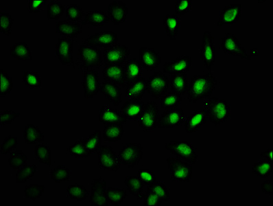
Immunofluorescence staining of Hela cells with CSB-PA345128LA01HU at 1:100, counter-stained with DAPI. The cells were fixed in 4% formaldehyde, permeabilized using 0.2% Triton X-100 and blocked in 10% normal Goat Serum. The cells were then incubated with the antibody overnight at 4°C. The secondary antibody was Alexa Fluor 488-congugated AffiniPure Goat Anti-Rabbit IgG(H+L).
Immunofluorescence staining of Hela cells with CSB-PA345128LA01HU at 1:100, counter-stained with DAPI. The cells were fixed in 4% formaldehyde, permeabilized using 0.2% Triton X-100 and blocked in 10% normal Goat Serum. The cells were then incubated with the antibody overnight at 4°C. The secondary antibody was Alexa Fluor 488-congugated AffiniPure Goat Anti-Rabbit IgG(H+L).
RAG2 Antibody
CSB-PA345128LA01HU
ApplicationsImmunoFluorescence, ELISA
Product group Antibodies
ReactivityHuman
TargetRAG2
Overview
- SupplierCusabio
- Product NameRAG2 Antibody
- Delivery Days Customer20
- ApplicationsImmunoFluorescence, ELISA
- CertificationResearch Use Only
- ClonalityPolyclonal
- ConjugateUnconjugated
- Gene ID5897
- Target nameRAG2
- Target descriptionrecombination activating 2
- Target synonymsRAG-2, V(D)J recombination-activating protein 2, recombination activating gene 2
- HostRabbit
- IsotypeIgG
- Protein IDP55895
- Protein NameV(D)J recombination-activating protein 2
- Scientific DescriptionCore component of the RAG complex, a multiprotein complex that mediates the DNA cleavage phase during V(D)J recombination. V(D)J recombination assembles a diverse repertoire of immunoglobulin and T-cell receptor genes in developing B and T-lymphocytes through rearrangement of different V (variable), in some cases D (diversity), and J (joining) gene segments. DNA cleavage by the RAG complex occurs in 2 steps: a first nick is introduced in the top strand immediately upstream of the heptamer, generating a 3-hydroxyl group that can attack the phosphodiester bond on the opposite strand in a direct transesterification reaction, thereby creating 4 DNA ends: 2 hairpin coding ends and 2 blunt, 5-phosphorylated ends. The chromatin structure plays an essential role in the V(D)J recombination reactions and the presence of histone H3 trimethylated at Lys-4 (H3K4me3) stimulates both the nicking and haipinning steps. The RAG complex also plays a role in pre-B cell allelic exclusion, a process leading to expression of a single immunoglobulin heavy chain allele to enforce clonality and monospecific recognition by the B-cell antigen receptor (BCR) expressed on individual B-lymphocytes. The introduction of DNA breaks by the RAG complex on one immunoglobulin allele induces ATM-dependent repositioning of the other allele to pericentromeric heterochromatin, preventing accessibility to the RAG complex and recombination of the second allele. In the RAG complex, RAG2 is not the catalytic component but is required for all known catalytic activities mediated by RAG1. It probably acts as a sensor of chromatin state that recruits the RAG complex to H3K4me3 (By similarity).
- ReactivityHuman
- Storage Instruction-20°C or -80°C
- UNSPSC41116161
Related products
Product group Antibodies
Anti-RAG2 Antibody144-62349
ApplicationsWestern Blot
ReactivityHuman, Mouse
TargetRAG2
- SizePrice
Product group Antibodies

RAG2 / RAG-2 AntibodyLS-C747587
ApplicationsWestern Blot
ReactivityHuman, Mouse
TargetRAG2
- SizePrice
Product group Antibodies
RAG2 Polyclonal AntibodyBS-6960R
ApplicationsImmunoFluorescence, ELISA, ImmunoCytoChemistry, ImmunoHistoChemistry, ImmunoHistoChemistry Frozen, ImmunoHistoChemistry Paraffin
ReactivityBovine, Canine, Equine, Human, Mouse, Porcine, Rabbit, Rat, Sheep
TargetRAG2
- SizePrice

Product group Antibodies
Anti-RAG2 Antibody Picoband(r)A00352-CARRIER-FREE
ApplicationsFlow Cytometry, Western Blot, ELISA
ReactivityHuman, Mouse
TargetRAG2
- SizePrice

Product group Antibodies
Anti-RAG2 AntibodyHPA065704
ApplicationsImmunoCytoChemistry
ReactivityHuman
TargetRAG2
- SizePrice
Product group Antibodies
RAG2 antibodyGTX109710
ApplicationsImmunoFluorescence, Western Blot, ImmunoCytoChemistry, ImmunoHistoChemistry, ImmunoHistoChemistry Paraffin
ReactivityHuman, Rat
TargetRAG2
- SizePrice
Product group Antibodies
ApplicationsWestern Blot, ELISA
ReactivityHuman
TargetRAG2
- SizePrice