


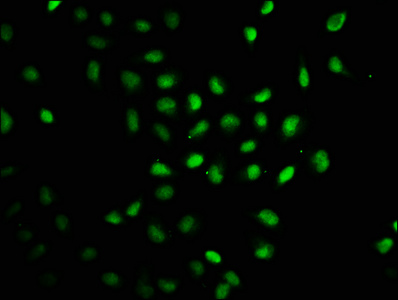
Immunofluorescent staining of human cell line REH shows localization to nucleoplasm.
Immunofluorescent staining of human cell line REH shows localization to nucleoplasm.
Anti-RAG2 Antibody
HPA065704
ApplicationsImmunoCytoChemistry
Product group Antibodies
ReactivityHuman
TargetRAG2
Overview
- SupplierAtlas Antibodies
- Product NameAnti-RAG2 Antibody
- Delivery Days Customer4
- ApplicationsImmunoCytoChemistry
- CertificationResearch Use Only
- ClonalityPolyclonal
- ConjugateUnconjugated
- Gene ID5897
- Target nameRAG2
- Target descriptionrecombination activating 2
- Target synonymsRAG-2, V(D)J recombination-activating protein 2, recombination activating gene 2
- HostRabbit
- IsotypeIgG
- Protein IDP55895
- Protein NameV(D)J recombination-activating protein 2
- Scientific DescriptionRecombinant Protein Epitope Signature Tag (PrEST) antigen sequence
- ReactivityHuman
- Storage Instruction-20°C,2°C to 8°C
- UNSPSC41116161
Datasheet
MSDS
Related products
Product group Antibodies
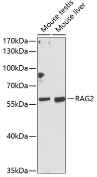
Anti-RAG2 Antibody144-62349
ApplicationsWestern Blot
ReactivityHuman, Mouse
TargetRAG2
- SizePrice
Product group Antibodies

RAG2 / RAG-2 AntibodyLS-C747587
ApplicationsWestern Blot
ReactivityHuman, Mouse
TargetRAG2
- SizePrice
Product group Antibodies
RAG2 Polyclonal AntibodyBS-6960R
ApplicationsImmunoFluorescence, ELISA, ImmunoCytoChemistry, ImmunoHistoChemistry, ImmunoHistoChemistry Frozen, ImmunoHistoChemistry Paraffin
ReactivityBovine, Canine, Equine, Human, Mouse, Porcine, Rabbit, Rat, Sheep
TargetRAG2
- SizePrice

Product group Antibodies
Anti-RAG2 Antibody Picoband(r)A00352-CARRIER-FREE
ApplicationsFlow Cytometry, Western Blot, ELISA
ReactivityHuman, Mouse
TargetRAG2
- SizePrice
Product group Antibodies
RAG2 AntibodyCSB-PA345128LA01HU
ApplicationsImmunoFluorescence, ELISA
ReactivityHuman
TargetRAG2
- SizePrice
Product group Antibodies
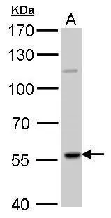
RAG2 antibodyGTX109710
ApplicationsImmunoFluorescence, Western Blot, ImmunoCytoChemistry, ImmunoHistoChemistry, ImmunoHistoChemistry Paraffin
ReactivityHuman, Rat
TargetRAG2
- SizePrice
Product group Antibodies
ApplicationsWestern Blot, ELISA
ReactivityHuman
TargetRAG2
- SizePrice