


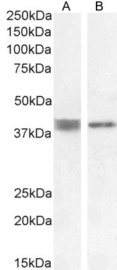
Figure 1. Western blot analysis of Renin receptor/ATP6AP2 using anti-Renin receptor/ATP6AP2 antibody (A02665-3). Electrophoresis was performed on a 5-20% SDS-PAGE gel at 70V (Stacking gel) / 90V (Resolving gel) for 2-3 hours. The sample well of each lane was loaded with 30 ug of sample under reducing conditions. Lane 1: human SiHa whole cell lysates, Lane 2: human HUVEC whole cell lysates, Lane 3: human U251 whole cell lysates, Lane 4: rat brain tissue lysates, Lane 5: rat heart tissue lysates, Lane 6: mouse brain tissue lysates, Lane 7: mouse heart tissue lysates. After electrophoresis, proteins were transferred to a nitrocellulose membrane at 150 mA for 50-90 minutes. Blocked the membrane with 5% non-fat milk/TBS for 1.5 hour at RT. The membrane was incubated with rabbit anti-Renin receptor/ATP6AP2 antigen affinity purified polyclonal antibody (Catalog # A02665-3) at 0.5 microg/mL overnight at 4°C, then washed with TBS-0.1%Tween 3 times with 5 minutes each and probed with a goat anti-rabbit IgG-HRP secondary antibody at a dilution of 1:5000 for 1.5 hour at RT. The signal is developed using an Enhanced Chemiluminescent detection (ECL) kit (Catalog # EK1002) with Tanon 5200 system. A specific band was detected for Renin receptor/ATP6AP2 at approximately 49 kDa. The expected band size for Renin receptor/ATP6AP2 is at 39 kDa.
. Renin receptor/ATP6AP2 was detected in an immunocytochemical section of HELA cells. Enzyme antigen retrieval was performed using IHC enzyme antigen retrieval reagent (AR0022) for 15 mins. The cells were blocked with 10% goat serum. And then incubated with 5 microg/mL rabbit anti-Renin receptor/ATP6AP2 Antibody (A02665-3) overnight at 4°C. DyLight®488 Conjugated Goat Anti-Rabbit IgG (BA1127) was used as secondary antibody at 1:500 dilution and incubated for 30 minutes at 37°C. The section was counterstained with DAPI. Visualize using a fluorescence microscope and filter sets appropriate for the label used.")
. Overlay histogram showing SiHa cells stained with A02665-3 (Blue line). To facilitate intracellular staining, cells were fixed with 4% paraformaldehyde and permeabilized with permeabilization buffer. The cells were blocked with 10% normal goat serum. And then incubated with rabbit anti-Renin receptor/ATP6AP2 Antibody (A02665-3, 1 microg/1x106 cells) for 30 min at 20°C. DyLight®488 conjugated goat anti-rabbit IgG (BA1127, 5-10 microg/1x106 cells) was used as secondary antibody for 30 minutes at 20°C. Isotype control antibody (Green line) was rabbit IgG (1 microg/1x106) used under the same conditions. Unlabelled sample without incubation with primary antibody and secondary antibody (Red line) was used as a blank control.")
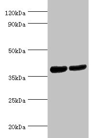
Figure 1. Western blot analysis of Renin receptor/ATP6AP2 using anti-Renin receptor/ATP6AP2 antibody (A02665-3). Electrophoresis was performed on a 5-20% SDS-PAGE gel at 70V (Stacking gel) / 90V (Resolving gel) for 2-3 hours. The sample well of each lane was loaded with 30 ug of sample under reducing conditions. Lane 1: human SiHa whole cell lysates, Lane 2: human HUVEC whole cell lysates, Lane 3: human U251 whole cell lysates, Lane 4: rat brain tissue lysates, Lane 5: rat heart tissue lysates, Lane 6: mouse brain tissue lysates, Lane 7: mouse heart tissue lysates. After electrophoresis, proteins were transferred to a nitrocellulose membrane at 150 mA for 50-90 minutes. Blocked the membrane with 5% non-fat milk/TBS for 1.5 hour at RT. The membrane was incubated with rabbit anti-Renin receptor/ATP6AP2 antigen affinity purified polyclonal antibody (Catalog # A02665-3) at 0.5 microg/mL overnight at 4°C, then washed with TBS-0.1%Tween 3 times with 5 minutes each and probed with a goat anti-rabbit IgG-HRP secondary antibody at a dilution of 1:5000 for 1.5 hour at RT. The signal is developed using an Enhanced Chemiluminescent detection (ECL) kit (Catalog # EK1002) with Tanon 5200 system. A specific band was detected for Renin receptor/ATP6AP2 at approximately 49 kDa. The expected band size for Renin receptor/ATP6AP2 is at 39 kDa.
Anti-Renin receptor/ATP6AP2 Antibody Picoband(r)
A02665-3-CARRIER-FREE
ApplicationsFlow Cytometry, ImmunoFluorescence, ImmunoPrecipitation, Western Blot, ImmunoCytoChemistry
Product group Antibodies
ReactivityHuman, Mouse, Rat
TargetATP6AP2
Overview
- SupplierBoster Bio
- Product NameAnti-Renin receptor/ATP6AP2 Antibody Picoband(r)
- Delivery Days Customer9
- ApplicationsFlow Cytometry, ImmunoFluorescence, ImmunoPrecipitation, Western Blot, ImmunoCytoChemistry
- CertificationResearch Use Only
- ClonalityPolyclonal
- Concentration500 ug/ml
- Gene ID10159
- Target nameATP6AP2
- Target descriptionATPase H+ transporting accessory protein 2
- Target synonyms(P)RR, APT6M8-9, ATP6IP2, ATP6M8-9, CDG2R, ELDF10, HT028, M8-9, MRXE, MRXSH, MSTP009, PRR, RENR, XMRE, XPDS, renin receptor, ATPase H(+)-transporting lysosomal-interacting protein 2, ATPase, H+ transporting, lysosomal (vacuolar proton pump) membrane sector associated protein M8-9, ATPase, H+ transporting, lysosomal accessory protein 2, ATPase, H+ transporting, lysosomal interacting protein 2, ER-localized type I transmembrane adapter, ER-localized type I transmembrane adaptor, N14F, V-ATPase M8.9 subunit, embryonic liver differentiation factor 10, prorenin receptor, renin/prorenin receptor, vacuolar ATP synthase membrane sector-associated protein M8-9, vacuolar proton ATP synthase membrane sector associated protein M8-9
- HostRabbit
- IsotypeIgG
- Protein IDO75787
- Protein NameRenin receptor
- Scientific DescriptionBoster Bio Anti-Renin receptor/ATP6AP2 Antibody Picoband® catalog # A02665-3. Tested in Flow Cytometry, IP, IF, ICC, WB applications. This antibody reacts with Human, Mouse, Rat. The brand Picoband indicates this is a premium antibody that guarantees superior quality, high affinity, and strong signals with minimal background in Western blot applications. Only our best-performing antibodies are designated as Picoband, ensuring unmatched performance.
- ReactivityHuman, Mouse, Rat
- Storage Instruction-20°C,2°C to 8°C
- UNSPSC12352203
Related products
Product group Antibodies
ApplicationsFlow Cytometry, Western Blot, ELISA
ReactivityHuman, Rat
- SizePrice
Product group Antibodies
Anti-ATP6AP2 Antibody144-06531
ApplicationsImmunoFluorescence, Western Blot
ReactivityHuman, Mouse, Rat
TargetATP6AP2
- SizePrice
Product group Antibodies
ATP6IP2 Polyclonal AntibodyBS-7691R
ApplicationsELISA, ImmunoHistoChemistry, ImmunoHistoChemistry Paraffin
ReactivityBovine, Canine, Chicken, Equine, Human, Mouse, Porcine, Rabbit, Rat
TargetATP6AP2
- SizePrice
Product group Antibodies
ATP6AP2 AntibodyCSB-PA002384ESR2HU
ApplicationsWestern Blot, ELISA, ImmunoHistoChemistry
ReactivityHuman, Mouse
TargetATP6AP2
- SizePrice
Product group Antibodies
References

ApplicationsFlow Cytometry, Western Blot, ELISA
ReactivityHuman, Rat
TargetATP6AP2
- SizePrice
Product group Antibodies
Atp6Ap2 Polyclonal AntibodyCAC07412
ApplicationsWestern Blot, ELISA
ReactivityMouse
TargetATP6AP2
- SizePrice
Product group Antibodies

ATP6AP2 / Renin Receptor AntibodyLS-C404725
ApplicationsELISA, ImmunoHistoChemistry
ReactivityHuman, Mouse, Rat
TargetATP6AP2
- SizePrice

Product group Antibodies
Anti-ATP6AP2 AntibodyHPA003156
ApplicationsWestern Blot, ImmunoHistoChemistry
ReactivityHuman
TargetATP6AP2
- SizePrice
![Renin Receptor antibody [N3C3] detects Renin Receptor protein by immunofluorescent analysis. Sample: DIV9 rat hippocampal neuron cells were fixed in 4% paraformaldehyde at RT for 15 min. Green: Renin Receptor stained by Renin Receptor antibody [N3C3] (GTX114169) diluted at 1:500. Red: Tau, an axon marker, stained by Tau antibody [GT287] (GTX634809) diluted at 1:500. Blue: Fluoroshield with DAPI (GTX30920).](https://www.genetex.com/upload/website/prouct_img/normal/GTX114169/GTX114169_43810_20211008_ICC_IF_R_w_23060501_403.webp)
Product group Antibodies
Renin Receptor antibody [N3C3]GTX114169
ApplicationsImmunoFluorescence, Western Blot, ImmunoCytoChemistry
ReactivityHuman, Mouse, Rat
TargetATP6AP2
- SizePrice
Product group Antibodies
Anti-ATP6AP2 AntibodyCAB6531
ApplicationsWestern Blot, ELISA
ReactivityHuman
TargetATP6AP2
- SizePrice