

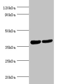
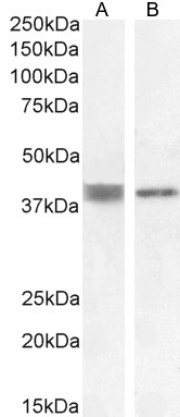
Western blot All lanes: Renin receptor antibody at 2ug/ml Lane 1: Hela whole cell lysate Lane 2: Mouse heart tissue Secondary Goat polyclonal to rabbit IgG at 1/10000 dilution Predicted band size: 40, 36 kDa Observed band size: 40 kDa

Western blot All lanes: Renin receptor antibody at 2ug/ml Lane 1: Hela whole cell lysate Lane 2: Mouse heart tissue Secondary Goat polyclonal to rabbit IgG at 1/10000 dilution Predicted band size: 40, 36 kDa Observed band size: 40 kDa
ATP6AP2 Antibody
CSB-PA002384ESR2HU
ApplicationsWestern Blot, ELISA, ImmunoHistoChemistry
Product group Antibodies
ReactivityHuman, Mouse
TargetATP6AP2
Overview
- SupplierCusabio
- Product NameATP6AP2 Antibody
- Delivery Days Customer20
- ApplicationsWestern Blot, ELISA, ImmunoHistoChemistry
- CertificationResearch Use Only
- ClonalityPolyclonal
- ConjugateUnconjugated
- Gene ID10159
- Target nameATP6AP2
- Target descriptionATPase H+ transporting accessory protein 2
- Target synonyms(P)RR, APT6M8-9, ATP6IP2, ATP6M8-9, CDG2R, ELDF10, HT028, M8-9, MRXE, MRXSH, MSTP009, PRR, RENR, XMRE, XPDS, renin receptor, ATPase H(+)-transporting lysosomal-interacting protein 2, ATPase, H+ transporting, lysosomal (vacuolar proton pump) membrane sector associated protein M8-9, ATPase, H+ transporting, lysosomal accessory protein 2, ATPase, H+ transporting, lysosomal interacting protein 2, ER-localized type I transmembrane adapter, ER-localized type I transmembrane adaptor, N14F, V-ATPase M8.9 subunit, embryonic liver differentiation factor 10, prorenin receptor, renin/prorenin receptor, vacuolar ATP synthase membrane sector-associated protein M8-9, vacuolar proton ATP synthase membrane sector associated protein M8-9
- HostRabbit
- IsotypeIgG
- Protein IDO75787
- Protein NameRenin receptor
- Scientific DescriptionFunctions as a renin and prorenin cellular receptor. May mediate renin-dependent cellular responses by activating ERK1 and ERK2. By increasing the catalytic efficiency of renin in AGT/angiotensinogen conversion to angiotensin I, it may also play a role in the renin-angiotensin system (RAS).1 Publication
- ReactivityHuman, Mouse
- Storage Instruction-20°C or -80°C
- UNSPSC41116161
Related products
Product group Antibodies
ApplicationsFlow Cytometry, Western Blot, ELISA
ReactivityHuman, Rat
- SizePrice

Product group Antibodies
Anti-Renin receptor/ATP6AP2 Antibody Picoband(r)A02665-3-CARRIER-FREE
ApplicationsFlow Cytometry, ImmunoFluorescence, ImmunoPrecipitation, Western Blot, ImmunoCytoChemistry
ReactivityHuman, Mouse, Rat
TargetATP6AP2
- SizePrice
Product group Antibodies
Anti-ATP6AP2 Antibody144-06531
ApplicationsImmunoFluorescence, Western Blot
ReactivityHuman, Mouse, Rat
TargetATP6AP2
- SizePrice
Product group Antibodies
ATP6IP2 Polyclonal AntibodyBS-7691R
ApplicationsELISA, ImmunoHistoChemistry, ImmunoHistoChemistry Paraffin
ReactivityBovine, Canine, Chicken, Equine, Human, Mouse, Porcine, Rabbit, Rat
TargetATP6AP2
- SizePrice
Product group Antibodies
References

ApplicationsFlow Cytometry, Western Blot, ELISA
ReactivityHuman, Rat
TargetATP6AP2
- SizePrice
Product group Antibodies
Atp6Ap2 Polyclonal AntibodyCAC07412
ApplicationsWestern Blot, ELISA
ReactivityMouse
TargetATP6AP2
- SizePrice
Product group Antibodies

ATP6AP2 / Renin Receptor AntibodyLS-C404725
ApplicationsELISA, ImmunoHistoChemistry
ReactivityHuman, Mouse, Rat
TargetATP6AP2
- SizePrice

Product group Antibodies
Anti-ATP6AP2 AntibodyHPA003156
ApplicationsWestern Blot, ImmunoHistoChemistry
ReactivityHuman
TargetATP6AP2
- SizePrice
![Renin Receptor antibody [N3C3] detects Renin Receptor protein by immunofluorescent analysis. Sample: DIV9 rat hippocampal neuron cells were fixed in 4% paraformaldehyde at RT for 15 min. Green: Renin Receptor stained by Renin Receptor antibody [N3C3] (GTX114169) diluted at 1:500. Red: Tau, an axon marker, stained by Tau antibody [GT287] (GTX634809) diluted at 1:500. Blue: Fluoroshield with DAPI (GTX30920).](https://www.genetex.com/upload/website/prouct_img/normal/GTX114169/GTX114169_43810_20211008_ICC_IF_R_w_23060501_403.webp)
Product group Antibodies
Renin Receptor antibody [N3C3]GTX114169
ApplicationsImmunoFluorescence, Western Blot, ImmunoCytoChemistry
ReactivityHuman, Mouse, Rat
TargetATP6AP2
- SizePrice
Product group Antibodies
Anti-ATP6AP2 AntibodyCAB6531
ApplicationsWestern Blot, ELISA
ReactivityHuman
TargetATP6AP2
- SizePrice