


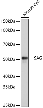
Figure 1. Western blot analysis of Retinal S antigen using anti-Retinal S antigen antibody (A03775). Electrophoresis was performed on a 5-20% SDS-PAGE gel at 70V (Stacking gel) / 90V (Resolving gel) for 2-3 hours. The sample well of each lane was loaded with 50ug of sample under reducing conditions. Lane 1: rat eye ball tissue lysates, Lane 2: mouse eye ball tissue lysates. After Electrophoresis, proteins were transferred to a Nitrocellulose membrane at 150mA for 50-90 minutes. Blocked the membrane with 5% Non-fat Milk/ TBS for 1.5 hour at RT. The membrane was incubated with rabbit anti-Retinal S antigen antigen affinity purified polyclonal antibody (Catalog # A03775) at 0.5 ug/mL overnight at 4 then washed with TBS-0.1%Tween 3 times with 5 minutes each and probed with a goat anti-rabbit IgG-HRP secondary antibody at a dilution of 1:10000 for 1.5 hour at RT. The signal is developed using an Enhanced Chemiluminescent detection (ECL) kit (Catalog # EK1002) with Tanon 5200 system. A specific band was detected for Retinal S antigen at approximately 45-55KD. The expected band size for Retinal S antigen is at 45KD.
Figure 1. Western blot analysis of Retinal S antigen using anti-Retinal S antigen antibody (A03775). Electrophoresis was performed on a 5-20% SDS-PAGE gel at 70V (Stacking gel) / 90V (Resolving gel) for 2-3 hours. The sample well of each lane was loaded with 50ug of sample under reducing conditions. Lane 1: rat eye ball tissue lysates, Lane 2: mouse eye ball tissue lysates. After Electrophoresis, proteins were transferred to a Nitrocellulose membrane at 150mA for 50-90 minutes. Blocked the membrane with 5% Non-fat Milk/ TBS for 1.5 hour at RT. The membrane was incubated with rabbit anti-Retinal S antigen antigen affinity purified polyclonal antibody (Catalog # A03775) at 0.5 ug/mL overnight at 4 then washed with TBS-0.1%Tween 3 times with 5 minutes each and probed with a goat anti-rabbit IgG-HRP secondary antibody at a dilution of 1:10000 for 1.5 hour at RT. The signal is developed using an Enhanced Chemiluminescent detection (ECL) kit (Catalog # EK1002) with Tanon 5200 system. A specific band was detected for Retinal S antigen at approximately 45-55KD. The expected band size for Retinal S antigen is at 45KD.
Anti-Retinal S antigen/SAG Antibody Picoband(r)
A03775-CARRIER-FREE
ApplicationsWestern Blot, ELISA
Product group Antibodies
ReactivityHuman, Mouse, Rat
TargetSAG
Overview
- SupplierBoster Bio
- Product NameAnti-Retinal S antigen/SAG Antibody Picoband(r)
- Delivery Days Customer9
- Application Supplier NoteTested Species: In-house tested species with positive results. Other applications have not been tested. Optimal dilutions should be determined by end users.
- ApplicationsWestern Blot, ELISA
- CertificationResearch Use Only
- ClonalityPolyclonal
- Concentration500 ug/ml
- Gene ID6295
- Target nameSAG
- Target descriptionS-antigen visual arrestin
- Target synonymsRP47, RP96, S-AG, S-arrestin, 48 kDa protein, S-antigen; retina and pineal gland (arrestin), arrestin 1, retinal S-antigen (48 KDa protein), rod arrestin, rod photoreceptor arrestin
- HostRabbit
- IsotypeIgG
- Protein IDP10523
- Protein NameS-arrestin
- Scientific DescriptionBoster Bio Anti-Retinal S antigen/SAG Antibody Picoband® catalog # A03775. Tested in ELISA, WB applications. This antibody reacts with Human, Mouse, Rat. The brand Picoband indicates this is a premium antibody that guarantees superior quality, high affinity, and strong signals with minimal background in Western blot applications. Only our best-performing antibodies are designated as Picoband, ensuring unmatched performance.
- ReactivityHuman, Mouse, Rat
- Storage Instruction-20°C,2°C to 8°C
- UNSPSC12352203
Related products
Product group Antibodies
SAG AntibodyCSB-PA020669YA01HU
ApplicationsELISA
ReactivityHuman
TargetSAG
- SizePrice
Product group Antibodies
ApplicationsWestern Blot, ImmunoHistoChemistry
ReactivityMouse, Rat
- SizePrice
Product group Antibodies

SAG / Arrestin AntibodyLS-C748112
ApplicationsWestern Blot
ReactivityHuman, Mouse, Rat
TargetSAG
- SizePrice

Product group Antibodies
Anti-SAG AntibodyHPA004946
ApplicationsImmunoHistoChemistry
ReactivityHuman
TargetSAG
- SizePrice
Product group Antibodies
ApplicationsImmunoPrecipitation, Western Blot, ImmunoCytoChemistry, ImmunoHistoChemistry
ReactivityMouse
TargetSAG
- SizePrice
Product group Antibodies
S-arrestin antibody [PDS-1]GTX40621
ApplicationsWestern Blot, ImmunoHistoChemistry, ImmunoHistoChemistry Frozen
ReactivityBovine, Human, Porcine, Rat
TargetSAG
- SizePrice
Product group Antibodies
Anti-SAG Antibody144-60158
ApplicationsWestern Blot
ReactivityHuman, Mouse, Rat
TargetSAG
- SizePrice