

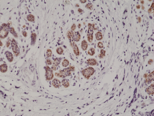
Immunohistochemical staining of formalin fixed and paraffin embedded human breast cancer tissue section using anti-S100B rabbit monoclonal antibody (Clone RM304) at a 1:1000 dilution.
Immunohistochemical staining of formalin fixed and paraffin embedded human breast cancer tissue section using anti-S100B rabbit monoclonal antibody (Clone RM304) at a 1:1000 dilution.
anti-S100-beta (human), Rabbit Monoclonal (RM304)
REV-31-1189-00
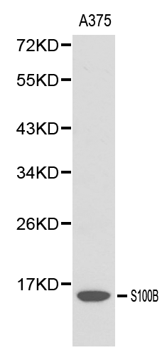
ApplicationsWestern Blot, ImmunoHistoChemistry
Product group Antibodies
ReactivityHuman
TargetS100B
Overview
- SupplierRevMAb Biosciences
- Product Nameanti-S100-beta (human), Rabbit Monoclonal (RM304)
- Delivery Days Customer2
- ApplicationsWestern Blot, ImmunoHistoChemistry
- CertificationResearch Use Only
- ClonalityMonoclonal
- Clone IDRM304
- Gene ID6285
- Target nameS100B
- Target descriptionS100 calcium binding protein B
- Target synonymsNEF, S100, S100-B, S100beta, protein S100-B, S-100 calcium-binding protein, beta chain, S-100 protein subunit beta, S100 calcium-binding protein, beta (neural)
- HostRabbit
- IsotypeIgG
- Protein IDP04271
- Protein NameProtein S100-B
- Scientific DescriptionRecombinant Antibody. This antibody reacts to human S100-B (S-100 protein beta chain). It may also react to mouse or rat S100-B, as predicted by immunogen homology. Applications: WB, IHC. Source: Rabbit. Liquid. 50% Glycerol/PBS with 1% BSA and 0.09% sodium azide. S100-beta (S100B) is a member of the S100 family of proteins containing 2 EF-hand calcium-binding motifs. S100 proteins are localized in the cytoplasm and/or nucleus of a wide range of cells and are involved in the regulation of a number of cellular processes such as cell cycle progression and differentiation. S100B may function in neurite extension, proliferation of melanoma cells, stimulation of Ca2+ fluxes, inhibition of PKC-mediated phosphorylation, astrocytosis and axonal proliferation, and inhibition of microtubule assembly. Chromosomal rearrangements and altered expression of this gene have been implicated in several diseases, including Alzheimers disease, Downs syndrome, epilepsy, amyotrophic lateral sclerosis, melanoma and type I diabetes. - S100-beta (S100B) is a member of the S100 family of proteins containing 2 EF-hand calcium-binding motifs. S100 proteins are localized in the cytoplasm and/or nucleus of a wide range of cells and are involved in the regulation of a number of cellular processes such as cell cycle progression and differentiation. S100B may function in neurite extension, proliferation of melanoma cells, stimulation of Ca2+ fluxes, inhibition of PKC-mediated phosphorylation, astrocytosis and axonal proliferation, and inhibition of microtubule assembly. Chromosomal rearrangements and altered expression of this gene have been implicated in several diseases, including Alzheimers disease, Downs syndrome, epilepsy, amyotrophic lateral sclerosis, melanoma and type I diabetes.
- ReactivityHuman
- Storage Instruction-20°C,2°C to 8°C
- UNSPSC41116161
Datasheet
Related products
Product group Antibodies
Anti-S100B AntibodyA28225
ApplicationsWestern Blot, ImmunoHistoChemistry
ReactivityHuman, Mouse, Rat
- SizePrice
Product group Antibodies

Anti-S100B [1C8]Ab02453-1.1
ApplicationsELISA
ReactivityHuman
TargetS100B
- SizePrice
Product group Antibodies
Anti-S100b Antibody130-00023
ApplicationsWestern Blot, ELISA
ReactivityHuman
- SizePrice

Product group Antibodies
Anti-S100B AntibodyAMAB91038
ApplicationsWestern Blot, ImmunoHistoChemistry
ReactivityHuman, Mouse, Rat
TargetS100B
- SizePrice
Product group Antibodies
References
S100B Polyclonal AntibodyBS-2015R
ApplicationsImmunoFluorescence, ELISA, ImmunoCytoChemistry, ImmunoHistoChemistry, ImmunoHistoChemistry Frozen, ImmunoHistoChemistry Paraffin
ReactivityHuman
TargetS100B
- SizePrice
Product group Antibodies

Goat anti-S100BEB12316
ApplicationsWestern Blot, ELISA, ImmunoHistoChemistry
ReactivityHuman
TargetS100B
- SizePrice
Product group Antibodies
S100B AntibodyCSB-PA020643ESR1HU
ApplicationsELISA, ImmunoHistoChemistry
ReactivityHuman
TargetS100B
- SizePrice
Product group Antibodies
ApplicationsImmunoPrecipitation, Western Blot, ImmunoCytoChemistry, ImmunoHistoChemistry
TargetS100B
- SizePrice

Product group Antibodies
References
ApplicationsImmunoFluorescence, ImmunoPrecipitation, Western Blot, ImmunoCytoChemistry, ImmunoHistoChemistry
ReactivityHuman, Mouse, Rat
TargetS100B
- SizePrice